
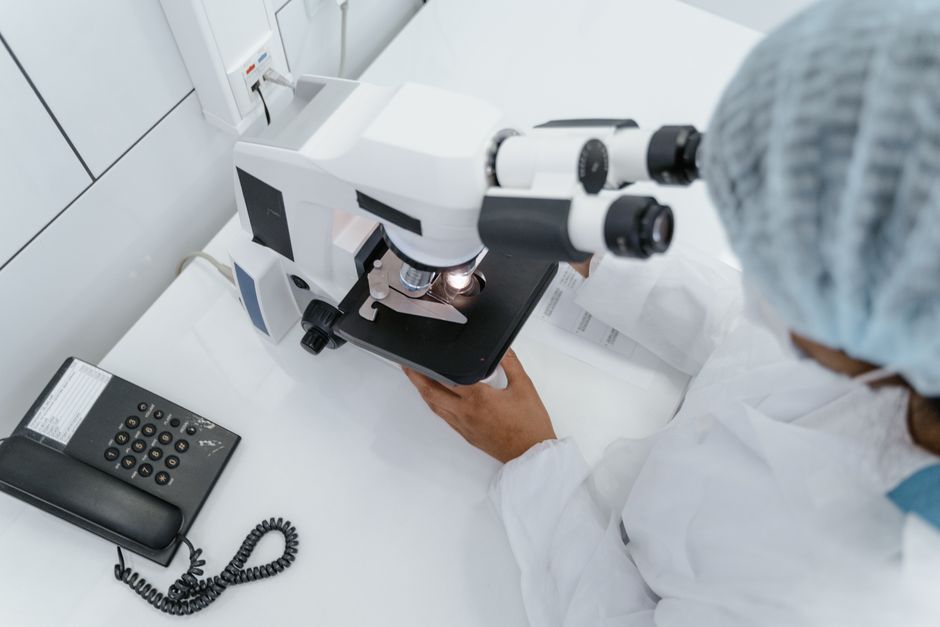
Super resolution microscopy is a set of imaging techniques that let scientists see structures smaller than the roughly 200-nanometer limit that ordinary light microscopes cannot cross. It works by using fluorescent molecules, lasers, and computer processing to either shrink the spot of light that illuminates a sample or to switch individual glowing molecules on and off one at a time. The result is a picture with detail far beyond what a standard light microscope can produce.
That 200-nanometer figure is not a flaw in any one instrument. It is a physical boundary. For most of the twentieth century, it was treated as the hard edge of what light could ever show us. Super resolution microscopy is the workaround that broke through it, and the scientists behind it won the 2014 Nobel Prize in Chemistry for making it work.
What Is Super Resolution Microscopy And How Does It Work?
Super resolution microscopy is any light-based imaging method that resolves details finer than the diffraction limit of light, which sits at roughly half the wavelength of the light being used — about 200 nanometers for visible light. It works by separating, in time or in space, fluorescent molecules that would otherwise blur together.
To understand why this is hard, picture two fireflies close together in the dark. From far away, their glow looks like one blob. You cannot tell there are two insects. The light from each one spreads out as it travels, and once the blobs overlap, no lens can pull them apart again. That spreading is diffraction.
Ernst Abbe described this limit mathematically in 1873. His formula showed that a microscope could not resolve two objects closer together than about half the light’s wavelength. For decades, this was taught as a law of nature.
The trick that beat it is clever. If you cannot separate two glowing molecules in space, you can separate them in time. Turn one on while the other stays dark, record its exact position, then switch them. Build up thousands of these snapshots and a computer stacks them into one sharp image. The blur is still there in each frame — but the center of each blur can be calculated precisely. Averaged over thousands of molecules, the picture becomes sharp.
Why Can’t Regular Microscopes See Very Small Things?
Regular light microscopes hit a wall set by physics, not engineering. When light passes through a lens, it bends and spreads. Even a perfect lens focuses light into a spot about 200 nanometers wide rather than a single point.
Anything smaller than that spot gets smeared out. Two structures 50 nanometers apart look like one fuzzy blob. This is the diffraction limit, and it applies to every microscope that uses visible light — no matter how expensive the lenses are.
Electron microscopes get around the limit by using electrons instead of light. Electrons have much shorter wavelengths, so they can resolve details down to fractions of a nanometer. But electron microscopy has real drawbacks for biology. Samples usually must be fixed, dehydrated, and placed in a vacuum, which kills living cells. It also struggles to label specific proteins with color the way fluorescent dyes can.
Super resolution microscopy sits in the middle. It offers resolution far better than standard light microscopy while keeping the ability to image specific molecules in cells, and in some methods, living cells.
What Are the Main Types of Super Resolution Microscopy?
Scientists group these techniques into two broad families. One family narrows the light itself. The other plays tricks with time and single molecules.
STED (stimulated emission depletion) belongs to the first family. It uses two laser beams. One excites fluorescent molecules in a spot. The second, shaped like a donut, immediately switches off the glow at the edges of that spot. What remains is a much smaller glowing point. Scanning this tiny point across the sample builds the image. STED can work in living cells and is relatively fast.
STORM and PALM belong to the second family. These methods switch individual fluorescent molecules on and off randomly, so only a sparse few glow at any moment. The microscope records the precise center of each glow, repeats this process thousands of times, and a computer assembles the final image. Resolution can reach tens of nanometers — well past the diffraction limit.
Structured illumination microscopy (SIM) takes a different route. It projects patterned light onto the sample and uses the way those patterns interact with the sample to extract finer detail mathematically. SIM offers less resolution gain than STED or STORM but works faster and with gentler light, which suits live-cell imaging.
- STED: narrows the light spot with a donut-shaped beam; good for live cells
- STORM/PALM: switches single molecules on and off over time; highest resolution
- SIM: uses patterned light and math; fastest and gentlest





How Do Scientists Actually Build These Images?
Every super resolution method depends on fluorescence. Scientists attach fluorescent tags — often antibodies or genetically encoded proteins — to the specific structures they want to see. When laser light hits a tag, it absorbs the energy and re-emits light at a different wavelength. That emitted light is what the camera captures.
The difference between methods lies in how they handle that light.
In STED, the donut beam does the work physically. It forces molecules at the edge of the spot to release their energy without emitting visible light, leaving only a tiny central region glowing. The microscope scans this point across the sample, line by line, like a printer building an image.
In STORM and PALM, the work happens computationally. Because only a few molecules glow at once, each one appears as an isolated blurry spot. Software calculates the exact center of each spot to a precision far better than the blur itself. After thousands of rounds, the software maps every position and produces the final image.
This is where a non-obvious point matters. The resolution gain in STORM and PALM is not optical. The microscope’s lenses are ordinary. The sharpness comes from math applied to thousands of individual detections. The physics of light still limits each single measurement — but averaging many measurements gets around it.
What Has Super Resolution Microscopy Revealed?
These techniques have changed how scientists understand the inside of cells. Before super resolution, many structures were simply invisible.
Researchers have used it to map how proteins cluster in cell membranes, how the cytoskeleton — the cell’s internal scaffolding — is organized, and how molecules move through synapses in the nervous system. It has helped reveal the structure of nuclear pores and the organization of mitochondria.
In neuroscience, super resolution imaging has shown that some synaptic proteins are arranged in precise patterns rather than scattered randomly. That kind of detail was not accessible with standard light microscopy.
The methods have also advanced microbiology. They have helped show how bacteria organize their internal structures, which was long assumed to be disorganized because bacteria are so small.
What these techniques have not done is replace electron microscopy or other methods. Each tool answers different questions. Super resolution is best when you need to see specific labeled molecules in their natural context, sometimes while the cell is still alive.
What Are the Limits and Challenges?
Super resolution microscopy is powerful, but it is not a magic window. Several real constraints apply.
First, resolution gains depend on the method. STORM and PALM can reach tens of nanometers. STED typically achieves somewhat less. SIM improves on the diffraction limit but by a smaller factor. No single number applies to all techniques.
Second, samples need bright, stable fluorescent labels. Not every molecule can be tagged effectively. If the tag does not work, the structure stays invisible.
Third, light can damage living cells. Higher laser power and longer imaging times increase the risk. This creates a trade-off: better resolution often means more light, which can harm the very cells being studied.
Fourth, these are complex, expensive instruments. They require trained operators, careful sample preparation, and significant computing power for image reconstruction. That limits how widely they are used outside well-funded labs.
Fifth, artifacts are a genuine concern. Poor labeling, sample movement, or errors in image processing can create structures that are not really there. Careful controls and validation matter.
Is Super Resolution Microscopy Used in Medicine?
Super resolution microscopy is primarily a research tool today, not a routine clinical test. It is used in laboratories to study disease mechanisms at the molecular level.
Researchers have applied it to study how proteins misfold in neurodegenerative conditions, how cancer cells reorganize their internal structures, and how pathogens interact with host cells. These findings inform our understanding of disease, though they are not yet standard diagnostic tools.
Some clinical applications are being explored. Pathologists have investigated whether super resolution methods could improve tissue analysis. But routine use in hospitals remains limited, and no clinical guidelines currently recommend these techniques for diagnosis. The evidence base for clinical utility is still developing.
It is worth being clear about this. The value of super resolution microscopy for basic science is well established. Its value as a clinical tool is not, at least not yet. Anyone claiming it is already transforming medical diagnosis is ahead of the evidence.
Frequently Asked Questions
What is the resolution limit of super resolution microscopy?
Super resolution methods can resolve details well below the roughly 200-nanometer diffraction limit of standard light microscopy. STORM and PALM can reach tens of nanometers, while STED and SIM achieve smaller gains.
Can super resolution microscopy image living cells?
Some methods, including STED and SIM, can image living cells, though higher laser power and longer imaging times raise the risk of light damage. STORM and PALM are more often used on fixed samples because they require many rounds of imaging.
How is super resolution microscopy different from electron microscopy?
Super resolution microscopy uses light and fluorescent labels, so it can show specific molecules in color and sometimes in living cells. Electron microscopy uses electrons for far higher resolution but usually requires fixed, dehydrated samples in a vacuum.
Who won the Nobel Prize for super resolution microscopy?
The 2014 Nobel Prize in Chemistry was awarded for the development of super resolution fluorescence microscopy. The prize recognized work that made it possible to see structures smaller than the diffraction limit.
