
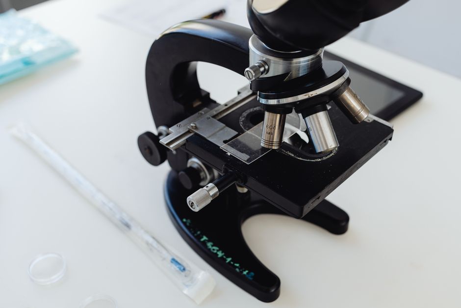
Microscopy is the use of instruments called microscopes to view objects and structures too small to be seen with the naked eye. The term covers a wide range of techniques, from simple light microscopes used in classrooms to electron microscopes that can resolve individual atoms. Microscopy is a foundational tool in medicine, biology, materials science, and forensics, and the type of microscope used depends entirely on what needs to be seen.
What Is Microscopy and How Does It Work?
Microscopy works by bending or focusing some form of energy — usually light or electrons — to produce a magnified image of a small object. The key principle is that the wavelength of the energy source determines how much detail can be resolved. Shorter wavelengths reveal smaller structures.
A light microscope uses visible light, which has a relatively long wavelength. That limits its useful magnification to roughly 1,000 to 1,500 times, and its resolving power to about 200 nanometers. Below that threshold, light waves blur together and no amount of magnification helps. This is not a limitation of engineering — it is a physical property of light itself.
Electron microscopes use a beam of electrons instead of light. Electrons have much shorter wavelengths, so they can resolve structures down to fractions of a nanometer. That is why electron microscopy can show the details of cell organelles, viruses, and even individual atoms in a crystal lattice.
Two numbers matter most in microscopy. Magnification is how much larger the image appears. Resolution is the smallest distance at which two points can still be distinguished as separate. High magnification without high resolution produces a large but blurry image. Resolution is the more important number.
What Are the Main Types of Microscopy?
There are several major categories, each suited to different samples and different questions. The list below covers the types most commonly used in clinical, research, and educational settings.
- Brightfield light microscopy — the standard microscope found in clinics and classrooms. Light passes through the sample and appears against a bright background. Best for stained or naturally pigmented specimens.
- Phase contrast microscopy — converts differences in refractive index into visible contrast. Useful for viewing living, unstained cells, which are nearly transparent under brightfield.
- Fluorescence microscopy — uses dyes or proteins that emit light when excited by a specific wavelength. Allows researchers to highlight specific molecules, structures, or cell types.
- Confocal microscopy — uses a pinhole to block out-of-focus light, producing sharp optical sections through thick samples. Common in cell biology and medical research.
- Electron microscopy — includes transmission electron microscopy (TEM) and scanning electron microscopy (SEM). TEM passes electrons through ultrathin samples to reveal internal structure. SEM scans a surface to produce detailed 3D-like images.
- Scanning probe microscopy — includes atomic force microscopy, which traces a physical probe across a surface to map it at the atomic level.
Each type has trade-offs. Light microscopy is fast, relatively inexpensive, and can be used on living samples. Electron microscopy offers far greater resolution but requires samples to be fixed, dehydrated, and placed in a vacuum — meaning nothing living survives the process.
What Is Microscopy Used For in Medicine?
Microscopy is central to medical diagnosis. When a pathologist examines a biopsy, they are using light microscopy to look at tissue structure, cell shape, and staining patterns. This is how most cancers are diagnosed and classified.
In hematology, a blood smear viewed under a microscope reveals the size, shape, and number of blood cells. This is how conditions like anemia, malaria, and certain leukemias are first identified. The microscope remains the reference standard for many of these assessments, even in an era of automated analyzers.
Microbiology relies on microscopy to identify bacteria, fungi, and parasites. Gram staining, for example, separates bacteria into two major groups based on cell wall structure — a distinction that has guided antibiotic selection for decades. While molecular testing has become more common, direct microscopic examination is still widely used because it is fast and does not require specialized equipment beyond a basic microscope.
In surgical settings, frozen section microscopy allows a pathologist to examine tissue during an operation and report back within minutes. This guides decisions about how much tissue to remove. It is one of the few diagnostic methods where speed directly affects the procedure itself.


How Is Microscopy Used Outside Medicine?
Microscopy is not limited to healthcare. Its applications span many fields.
In materials science, microscopy is used to inspect metals, ceramics, and polymers for defects, grain structure, and failure points. A scanning electron microscope can reveal cracks or impurities that would be invisible otherwise. Semiconductor manufacturing depends on electron microscopy to inspect circuit features measured in nanometers.
In forensic science, microscopy helps analyze trace evidence such as fibers, hair, paint chips, and gunshot residue. The comparison microscope, which views two specimens side by side, is a standard tool in crime labs.
Environmental scientists use microscopy to identify airborne particles, study soil composition, and monitor water quality. Pollen analysis, asbestos detection, and microplastic identification all rely on microscopic examination.
In food science, microscopy is used to detect contaminants, study food structure, and verify labeling claims. It also plays a role in agricultural research, where plant tissues and pests are examined routinely.
What Is the Difference Between Light and Electron Microscopy?
The differences come down to resolution, sample preparation, and what each method can realistically show.
| Feature | Light Microscopy | Electron Microscopy |
|---|---|---|
| Energy source | Visible light | Electron beam |
| Resolution limit | About 200 nanometers | Below 1 nanometer |
| Magnification range | Up to about 1,500x | Up to 1,000,000x or more |
| Living samples | Yes | No |
| Sample preparation | Minimal to moderate | Extensive (fixed, dehydrated, vacuum) |
| Cost and complexity | Low to moderate | High |
| Color information | Yes (with stains) | No (images are grayscale, often false-colored) |
The practical takeaway: if you need to see living cells or get a quick answer, light microscopy is the tool. If you need to see the internal structure of a virus or the arrangement of atoms, electron microscopy is required. Neither replaces the other.
What Are the Limitations of Microscopy?
Every microscopy method has constraints. Recognizing them matters because a microscope image is not a perfect replica of reality — it is an interpretation shaped by the instrument and the preparation method.
Sample preparation can introduce artifacts. When tissue is fixed, sliced, and stained, the process can alter structures. What appears under the microscope may not look exactly the same in the living body. Experienced pathologists learn to recognize common artifacts, but they remain a source of diagnostic uncertainty.
Resolution limits are absolute for each method. No light microscope can resolve two objects closer than roughly 200 nanometers apart, regardless of how good the lenses are. Super-resolution fluorescence techniques have pushed past this limit in research settings, but they require specialized equipment and are not routine in clinical labs.
Electron microscopy, despite its power, cannot be used on living tissue. The vacuum environment and the heavy metal stains used to create contrast kill any biological activity. This means electron microscopy provides a snapshot, not a live view.
Cost and access also matter. Electron microscopes cost hundreds of thousands of dollars and require trained operators and dedicated facilities. Light microscopes are affordable and widely available. This gap shapes where and how each technique is used.
How Has Microscopy Changed Over Time?
The first compound microscopes appeared in the late 16th century, and by the 17th century, researchers like Antonie van Leeuwenhoek were using single-lens microscopes to observe bacteria and protozoa for the first time. Those early instruments could magnify around 200 to 300 times.
Over the following centuries, lens quality improved, staining techniques were developed, and the theoretical limits of light microscopy were worked out. The invention of the electron microscope in the 1930s broke through the resolution barrier that light had imposed, opening up a new scale of observation.
More recently, digital imaging has transformed how microscopy is practiced. Cameras, software, and automated analysis now allow rapid scanning of slides, remote consultation between pathologists, and the use of artificial intelligence to flag suspicious regions. These tools do not replace the microscope — they extend what can be done with it.
One non-obvious point: the human eye is still part of the optical system in most clinical microscopy. The quality of the final image depends not just on the lenses and the camera but on the training and attention of the person looking through the eyepiece. Automated systems are improving, but for many diagnostic tasks, the pathologist’s eye remains the final arbiter.
Frequently Asked Questions
What is microscopy in simple terms?
Microscopy is the practice of using a microscope to see objects too small for the naked eye. It includes both the instruments and the techniques used to prepare and view samples.
What are the four main types of microscopy?
The four most commonly referenced types are brightfield light microscopy, phase contrast microscopy, fluorescence microscopy, and electron microscopy. Each is suited to different samples and levels of detail.
What is the difference between magnification and resolution?
Magnification makes an image larger, while resolution determines how much fine detail can actually be distinguished. High magnification without good resolution just produces a bigger blurry image.
Can you see living cells with a microscope?
Yes, with light microscopy you can view living cells, especially using phase contrast or similar techniques. Electron microscopy cannot be used on living samples because it requires a vacuum and fixed tissue.
