
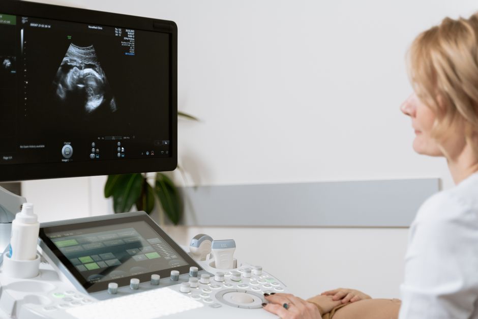
Fetal biometry is the part of an ultrasound exam where the sonographer measures the baby’s head, belly, and thigh bone. Those numbers are compared against growth charts for babies at the same gestational age. The result tells your clinician whether your baby is measuring small, average, or large, and whether growth is tracking normally over time.
It is one of the most routine parts of prenatal care. It is also one of the most misunderstood, partly because a single set of measurements can look alarming when it is actually normal variation.
What Is Fetal Biometry Ultrasound Measurements Explained?
Fetal biometry is a set of standardized measurements taken during an obstetric ultrasound to estimate fetal size and growth. The sonographer uses the machine’s calipers to measure specific bony and soft-tissue landmarks, and the ultrasound software converts those measurements into an estimated fetal weight.
The core measurements are:
- Biparietal diameter (BPD) — the width of the skull from side to side
- Head circumference (HC) — the distance around the skull
- Abdominal circumference (AC) — the distance around the baby’s belly
- Femur length (FL) — the length of the thigh bone
Some protocols also include humerus length, transcerebellar diameter, or other measurements depending on what the clinician is looking for. The four above are the standard set used to calculate estimated fetal weight.
Each measurement is plotted on a growth curve. The curve shows how a typical fetus at that exact gestational age compares. Your baby’s measurement is expressed as a percentile — for example, the 50th percentile means half of fetuses at that age measure larger and half measure smaller.
How Accurate Is Estimated Fetal Weight?
Estimated fetal weight from ultrasound is a good tool, not a precise scale. Research consistently shows it carries a margin of error of roughly 10 to 15 percent in either direction, and that margin widens at the extremes — very small or very large babies.
This matters because a baby estimated at 3,500 grams could actually weigh anywhere from about 3,000 to 4,000 grams. That range spans what clinicians would call normal and what they might call large.
The abdominal circumference is the single measurement most strongly tied to estimated fetal weight, which is why it gets extra attention when growth is being assessed. But AC is also the measurement most affected by fetal position, breathing movements, and how the calipers are placed. A wiggly baby or an awkward angle can shift the number.
Ultrasound machines use formulas developed from population data — most commonly the Hadlock formulas — to convert measurements into a weight estimate. Those formulas were built on specific populations and perform somewhat differently across different groups. This is one reason clinicians look at trends over multiple scans rather than reacting to a single number.
What Do the Percentiles Mean?
A percentile tells you where your baby’s measurement falls compared to other fetuses at the same gestational age. The 50th percentile is the middle. The 10th percentile means 10 percent of fetuses at that age measure smaller, and 90 percent measure larger.
Clinicians generally flag measurements below the 10th percentile or above the 90th percentile for closer attention. These cutoffs are widely used in obstetric practice, though they are screening thresholds, not diagnoses.
Here is where a lot of confusion happens. A baby at the 15th percentile is not “failing to grow.” A baby at the 85th percentile is not necessarily “too big.” Many perfectly healthy babies sit at the edges of the curve simply because they are genetically smaller or larger — just as adults vary in height and build.
Parental size matters. A baby born to two small parents is expected to measure smaller. A baby born to two large parents is expected to measure larger. A clinician who flags a low percentile will usually ask about parental height and weight, previous babies’ birth weights, and ethnic background before drawing conclusions.
The trend matters more than any single snapshot. A baby consistently tracking along the 20th percentile is usually a different situation from a baby who was at the 50th percentile and dropped to the 20th between scans.
When Is Fetal Biometry Measured?
Biometry is typically performed at the anatomy scan, usually between 18 and 22 weeks, and again in the third trimester if growth monitoring is needed. The timing and frequency depend on your individual pregnancy and your clinician’s judgment.
Earlier in pregnancy, biometry is most useful for dating. Crown-rump length in the first trimester is the most accurate way to establish gestational age, and it is most reliable when done early. Later scans are less precise for dating because babies of the same age vary more in size as pregnancy progresses.
In the third trimester, biometry is used to assess growth. Clinicians may recommend additional growth scans if there are risk factors such as high blood pressure, diabetes, or a prior small baby — or if fundal height measurements (the tape measure across your belly) suggest a discrepancy.
Serial scans — repeated measurements over weeks — are generally more informative than a single scan. They allow the clinician to see whether growth is following a normal curve or deviating from it.
What Does It Mean If My Baby Measures Small?
A small measurement does not automatically mean something is wrong. Many small babies are constitutionally small — healthy, just genetically on the smaller side — and are born at low birth weight without any complications.
When a fetus measures below the 10th percentile, clinicians use the term fetal growth restriction (FGR) only when there is evidence the fetus is not meeting its growth potential. That distinction requires more than one measurement. It involves looking at the trend, amniotic fluid volume, umbilical artery blood flow on Doppler ultrasound, and other factors.
True growth restriction happens when the placenta is not delivering enough oxygen and nutrients. This can be caused by conditions like preeclampsia, chronic hypertension, or placental problems. It requires monitoring and sometimes early delivery, depending on severity and gestational age.
The distinction between a small-but-healthy baby and a growth-restricted baby is one of the harder calls in obstetrics. This is why a low percentile on one scan usually leads to repeat scanning rather than immediate action.
What Does It Mean If My Baby Measures Large?
A large measurement — typically above the 90th percentile — is called macrosomia when suspected. Like small measurements, large ones are often just normal variation in bigger families.
The main clinical concern with suspected macrosomia is shoulder dystocia, a delivery complication where the baby’s shoulder gets stuck behind the pubic bone. This is uncommon, and ultrasound estimates of large babies are notably imprecise. Research has found that ultrasound tends to overestimate weight in large fetuses more than it underestimates it.
This imprecision is why suspected macrosomia alone rarely drives decisions about delivery mode. Clinicians weigh estimated weight alongside maternal factors like diabetes, prior delivery history, and pelvic shape.
Gestational diabetes is a common reason for larger-than-expected measurements, because higher maternal blood sugar can drive fetal growth. Managing blood sugar is the main lever in those cases.
What Biometry Cannot Tell You
Biometry estimates size. It does not diagnose chromosomal conditions, structural abnormalities, or genetic syndromes. Those require different assessments — detailed anatomy scanning, genetic screening, or diagnostic testing.
Biometry also cannot predict a baby’s future height, intelligence, or health outcomes. A baby at the 30th percentile at 32 weeks is not destined to be a small child or adult.
Perhaps the most useful thing to understand is that biometry is a screening tool. It flags patterns that deserve attention. It does not deliver verdicts. The clinical picture — your health, your history, the trend across scans, and other findings — is what turns a number into a meaningful assessment.
If a measurement concerns you, ask your clinician three questions: What percentile is this? How does it compare to my previous scan? And what is the plan for following up? Those answers will tell you far more than the raw number alone.
Frequently Asked Questions
How accurate is ultrasound estimated fetal weight?
Ultrasound estimated fetal weight is typically accurate within about 10 to 15 percent in either direction. Accuracy is lower at the extremes of very small or very large babies.
What percentile is considered normal in fetal biometry?
Measurements between the 10th and 90th percentiles are generally considered within the normal range. Values outside that range are usually flagged for closer monitoring rather than treated as abnormal on their own.
Does a low percentile mean my baby has growth restriction?
No. A single low percentile does not confirm growth restriction, which requires evidence the fetus is not meeting its growth potential. Clinicians typically look at trends across scans, blood flow studies, and other factors before making that call.
Can fetal biometry tell the sex of the baby?
No. Fetal biometry measures size and growth, not sex. Sex is determined by directly visualizing the genitalia during ultrasound, usually at the anatomy scan.