
Diagnostic medical imaging is the use of machines to look inside the body without surgery. It helps doctors find disease, check how well organs work, and see whether treatment is helping. The main types are X-ray, CT, MRI, ultrasound, and nuclear medicine, including PET scans. Each uses different physics and shows different things.
Imaging has changed medicine deeply. A broken bone that once needed exploratory surgery can now be seen in minutes. A tumor too small to feel can be found on a scan. But imaging also has limits, costs, and risks. Understanding what each type does — and does not do — helps you ask better questions when your doctor orders a scan.
What Is Diagnostic Medical Imaging and How Does It Work?
Diagnostic imaging creates pictures of the inside of the body using energy or sound waves. The image lets a doctor see structure, and sometimes function, without cutting into the body.
The physics matter because they explain why one scan shows bone clearly and another shows soft tissue. X-rays pass through the body and are absorbed differently by dense tissue like bone and by air in the lungs. The pattern that reaches a detector becomes an image. CT uses the same X-ray energy but takes many pictures from different angles and combines them by computer into cross-sections.
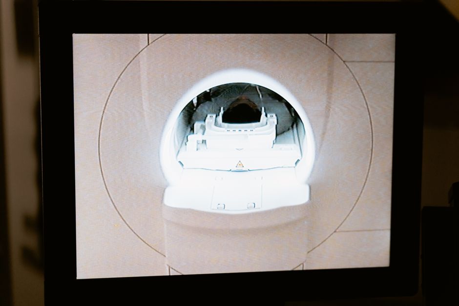
MRI uses strong magnetic fields and radio waves. It does not use ionizing radiation. It works because hydrogen atoms in water and fat respond to the magnetic field in ways that differ by tissue. That is why MRI is so good at showing soft tissue like the brain, spinal cord, and joints.
Ultrasound uses high-frequency sound waves that bounce off tissue. A computer turns the echoes into an image. Nuclear medicine works differently again: a small amount of a radioactive substance is given, usually by injection, and the scanner detects where it collects. PET scans are a type of nuclear medicine that show metabolic activity — how actively cells are using energy.
One clarification that surprises many people: the word “diagnostic” does not mean the scan makes the diagnosis. The scan produces images. A trained radiologist interprets them, and the referring doctor puts that together with your history, exam, and other tests. Imaging is one piece of a larger picture.
What Are the Main Types of Diagnostic Medical Imaging?
Five categories cover most diagnostic imaging. They differ in what they show, how long they take, and what risks they carry.
- X-ray — fast, low cost, best for bone and chest. Uses a small dose of ionizing radiation.
- CT (computed tomography) — detailed cross-sections, excellent for trauma, chest, and abdomen. Uses ionizing radiation, more than a single X-ray.
- MRI (magnetic resonance imaging) — excellent soft-tissue detail, no ionizing radiation. Takes longer and can be uncomfortable for people who dislike enclosed spaces.
- Ultrasound — real-time images, no ionizing radiation, widely used in pregnancy and for the heart and abdomen. Quality depends on the operator.
- Nuclear medicine and PET — shows function and activity, useful in cancer staging and some heart and brain conditions. Involves a radioactive tracer.
Each type has strengths and trade-offs. A CT can show a kidney stone in minutes; an MRI would be slower and often unnecessary for that question. An MRI can show a torn ligament that an X-ray cannot see at all. The right choice depends on the question being asked.
How Do Doctors Decide Which Imaging Test to Use?
Doctors choose imaging based on the clinical question, the body part, the urgency, and the person’s individual risks. There is rarely one “best” scan in the abstract.
The first factor is what needs to be seen. Bone and lung problems often call for X-ray or CT. Soft tissue in the brain, spine, or joints often calls for MRI. Pregnancy and many abdominal questions often call for ultrasound first because it avoids ionizing radiation.
Urgency matters too. In an emergency, speed and availability may outweigh other considerations. A CT scan in a trauma bay can find internal bleeding in minutes. MRI, which can take much longer, is not practical in that setting.
Individual factors also shape the decision. Radiation exposure accumulates over a lifetime, so doctors weigh whether a scan using ionizing radiation is truly needed, especially in younger people and in pregnancy. Kidney function matters for CT scans that use iodine-based contrast dye. Metal implants and some medical devices can make MRI unsafe or limit its use, so patients are screened beforehand.
Many health systems use evidence-based guidelines, often called appropriateness criteria, to help doctors pick the right test. These guidelines are developed by specialty societies and are updated as evidence changes. They are a useful reference, not a rulebook that overrides clinical judgment.
What Are the Risks and Safety Considerations?
The main safety consideration with X-ray and CT is ionizing radiation. The dose from a single X-ray is very small. CT scans use higher doses, and the amount varies widely by body part and protocol.
Radiation risk is real but should be understood in proportion. At the doses used in diagnostic imaging, any added cancer risk is small for an individual. The concern is cumulative — many scans over a lifetime add up. That is why the guiding principle in radiology is to justify every scan and use the lowest dose that answers the question. This principle is called ALARA, for “as low as reasonably achievable.”
MRI does not use ionizing radiation, which is a genuine advantage. But it is not risk-free. The strong magnetic field can affect certain implants and devices, and the scanner is loud and enclosed. Some people need sedation or an open MRI. Contrast agents used in MRI, which contain gadolinium, carry their own considerations, and clinicians weigh these case by case.
Ultrasound is generally considered very safe because it uses sound waves, not radiation. It is the standard first-line imaging method in pregnancy. As with any test, it should be used when there is a clinical reason.
Nuclear medicine and PET involve a radioactive tracer. The amount is small and clears from the body over time, but the risks and benefits should be discussed with the referring doctor, especially in pregnancy and breastfeeding.
If you are pregnant or think you might be, tell the imaging staff before any scan. This is one of the most important things you can do. Staff can then adjust the approach or confirm with your doctor whether the scan is needed.
How Should You Prepare for an Imaging Appointment?
Preparation depends on the type of scan. Some need no preparation at all. Others require fasting, holding certain medications, or arriving early.
For many CT scans with contrast, you may be asked not to eat for a few hours beforehand. For some abdominal ultrasounds, you may need a full bladder or to fast. For MRI, you will be asked about implants, pacemakers, and metal in your body. Remove jewelry and metal objects before the scan.
Tell the staff about allergies, especially to contrast dye, and about kidney problems. Tell them if you are pregnant or breastfeeding. Bring a list of your medications and any prior imaging studies if you have them.
During the scan, you may need to lie still. Some scans ask you to hold your breath for short periods. If you feel anxious in enclosed spaces, say so in advance — staff can often help.
What Happens After an Imaging Test?
A radiologist reviews the images and writes a report. Your referring doctor then interprets that report in the context of your symptoms, exam, and other tests.
Timing varies. Some results are available the same day; others take longer. Ask when and how you will get your results.
One point worth knowing: a scan can find something that turns out not to matter — an incidental finding. These are common, especially on CT and MRI. Many are harmless, but some need follow-up. Your doctor will help decide whether further testing is needed. This is one reason imaging is not a substitute for clinical judgment.
Does Imaging Replace a Physical Exam or Other Tests?
No. Imaging adds information; it does not replace history-taking, physical examination, or laboratory tests. Each answers different questions.
A physical exam can reveal tenderness, swelling, or a mass. Lab tests can show infection, organ function, or markers of disease. Imaging shows structure and, in some cases, function. Doctors combine these sources because no single test tells the whole story.
Imaging is also not a screening tool for everything. Screening programs exist for specific conditions, such as certain cancers, and are based on evidence about who benefits. Using imaging broadly without a clinical reason can lead to unnecessary findings, anxiety, and further procedures.
Frequently Asked Questions
What is the difference between diagnostic and screening imaging?
Diagnostic imaging is done to investigate a specific symptom or question, while screening imaging looks for disease in people without symptoms. Screening programs are limited to conditions where evidence shows a clear benefit.
Is MRI safer than CT because it has no radiation?
MRI avoids ionizing radiation, which is a real advantage, but it still has risks from strong magnetic fields and contrast agents. The safer choice depends on the clinical question and the person’s individual circumstances.
How long does a diagnostic imaging test take?
Times vary widely: an X-ray takes minutes, a CT scan usually a few minutes, and an MRI can take 30 minutes or longer. Ultrasound time depends on what is being examined.
Can I refuse an imaging test my doctor recommends?
You can discuss any recommended test with your doctor, including its risks, benefits, and alternatives. Shared decision-making is appropriate, though in emergencies some scans may be time-critical.
