
Computed tomography, commonly called a CT scan, is a medical imaging technique that uses X-rays and computer processing to create detailed cross-sectional pictures of the body. Unlike a standard X-ray, which flattens the body into a single two-dimensional image, a CT scan takes many X-ray images from different angles and combines them to show slices of bones, blood vessels, and soft tissues. This technology allows doctors to see the size, shape, and position of structures inside the body with far more clarity than a traditional X-ray.
What Is Computed Tomography and How Does It Work?
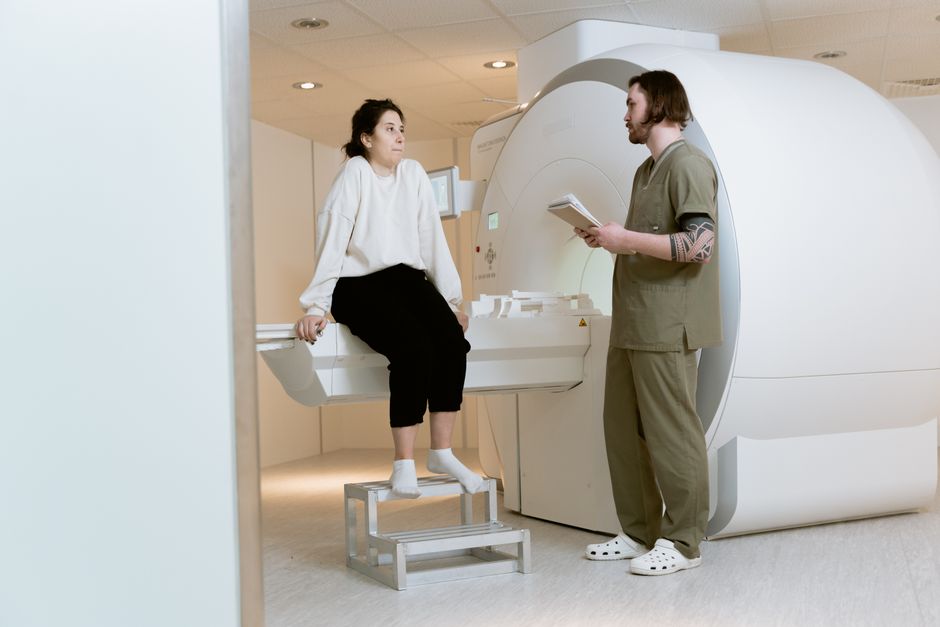
A CT scanner is a large, donut-shaped machine. You lie on a table that slides through the center opening while the X-ray tube rotates around you. The detector on the opposite side captures the X-rays that pass through your body.
The key is in the math. The scanner takes hundreds of individual X-ray projections during each rotation. A computer then uses those projections to reconstruct a detailed cross-sectional image, known as a slice. Modern scanners can capture these slices in less than a second and stack them to create a three-dimensional view of the area being examined.
The images appear in shades of gray. Dense materials like bone appear white because they block most X-rays. Soft tissues like the liver, kidneys, and muscles appear in varying shades of gray because they allow different amounts of radiation through. Air appears black. This contrast is what lets radiologists identify abnormalities like tumors, blood clots, or fractures.
Why Would a Doctor Order a CT Scan?
CT scans are used for a wide range of diagnostic purposes. They are often the fastest way to evaluate a patient in an emergency setting because they are quick and highly accurate.
Common reasons for a CT scan include:
- Evaluating trauma after a car accident or fall to check for internal bleeding or fractures
- Detecting and monitoring tumors or cancers in the chest, abdomen, or pelvis
- Diagnosing blood clots in the lungs, known as pulmonary embolism
- Identifying conditions like appendicitis, kidney stones, or diverticulitis
- Guiding biopsies or other minimally invasive procedures
- Assessing the severity of stroke or other brain conditions
CT is particularly valuable in emergency medicine because it can quickly rule out life-threatening conditions. A scan of the head takes only a few minutes and can clearly show bleeding in the brain after an injury. A scan of the chest can rapidly confirm or exclude a large pulmonary embolism.
CT Scan vs. MRI: What Is the Difference?
Many people confuse CT scans with magnetic resonance imaging, or MRI. They are fundamentally different technologies with different strengths.
A CT scan uses X-ray radiation. It is excellent at showing bone, dense structures, and acute bleeding. It is fast, usually taking less than 10 minutes from start to finish. It is the preferred test in emergencies and for evaluating the chest, abdomen, and pelvis.
An MRI uses a powerful magnetic field and radio waves. It does not use ionizing radiation. It provides superior contrast for soft tissues like the brain, spinal cord, ligaments, and cartilage. An MRI takes longer, often 30 to 60 minutes, and requires the patient to lie still inside a narrow tube.
The choice between CT and MRI depends on what the doctor is looking for. For a suspected kidney stone, a CT scan is the standard. For a torn knee ligament, an MRI is the better test. For evaluating the brain, both are used, but MRI generally shows soft tissue detail better while CT is faster and better for detecting acute bleeding.





What Are the Risks of Radiation Exposure?
CT scans do expose you to ionizing radiation. This is the main risk associated with the procedure. The amount of radiation varies depending on the body part being scanned and the machine settings.
The radiation dose from a CT scan is higher than a standard X-ray. For example, a chest X-ray delivers a very small dose, while a CT scan of the abdomen delivers more. The exact numbers depend on the equipment and protocol, but the general principle is consistent: more radiation exposure carries a small increase in the lifetime risk of cancer.
This risk is real but small. For a single CT scan, the estimated increase in lifetime cancer risk is very low. The medical benefit of an accurate, timely diagnosis almost always outweighs this small risk. Doctors are trained to order CT scans only when the information they provide will change patient management.
Children are more sensitive to radiation than adults. Their bodies are still developing, and they have more years of life ahead in which radiation-related effects could appear. For this reason, pediatric CT protocols use lower radiation doses, and doctors are more cautious about ordering CT scans in children. If a child needs a CT scan, ask the facility whether they use pediatric-specific settings.
If you are pregnant or think you might be, tell your doctor before the scan. The developing fetus is sensitive to radiation. In most cases, alternative imaging like ultrasound or MRI is preferred during pregnancy. However, in a life-threatening emergency, the benefit of a CT scan may still outweigh the risk to the fetus.
What Happens During a CT Scan?
The experience is straightforward. You will be asked to remove metal objects like jewelry, glasses, and dentures because they interfere with the images. You may be given a hospital gown to wear.
Depending on the type of scan, you may receive contrast material. This is often called a “dye,” though it is not a dye in the coloring sense. It is a liquid that contains iodine, and it makes certain structures appear brighter on the images. It can be given through an IV in your arm, swallowed as a drink, or given as an enema depending on the area being examined.
During the scan, you will lie on the table. The table moves slowly through the scanner. You will hear whirring and clicking noises. The technologist will be in an adjacent room watching you through a window and communicating through an intercom. You will be asked to hold your breath for short periods to prevent motion blur on the images.
The scan itself is painless. The entire process, including setup, typically takes 15 to 30 minutes. The actual scanning time is often less than a few minutes.
What Are the Side Effects of Contrast Material?
Most people who receive IV contrast feel a warm flush that spreads through the body. Some people notice a metallic taste in their mouth. These sensations are normal and fade within a minute or two.
Allergic reactions to contrast material are possible but uncommon. Mild reactions include hives or itching. Severe reactions, such as difficulty breathing or a drop in blood pressure, are rare. Let the technologist know immediately if you feel any discomfort during the contrast injection.
People with kidney disease need special consideration. The iodine-based contrast material is cleared from the body by the kidneys. In people with significantly reduced kidney function, contrast can cause a further decline called contrast-induced nephropathy. Your doctor may order a blood test to check your kidney function before giving IV contrast, especially if you have diabetes or known kidney problems.
If you take metformin, a common diabetes medication, your doctor may ask you to stop it temporarily before receiving contrast. This is a precaution because combining metformin with contrast can, in rare cases, lead to a dangerous condition called lactic acidosis, particularly in people with kidney impairment.
How Should You Prepare for a CT Scan?
Preparation depends on the type of scan you are having. For a head or limb scan, no special preparation is needed. For a scan of the abdomen or pelvis, your doctor may ask you to fast for several hours beforehand.
If you are having oral contrast, you will be given specific instructions about when to drink it. This usually involves drinking the contrast liquid over a period of time before the scan so it can coat your digestive tract.
Tell your doctor about all medications you take, including over-the-counter drugs and supplements. Mention any allergies, especially to iodine or shellfish. Having a shellfish allergy does not automatically mean you will react to contrast material, but your doctor needs to know about it.
Wear comfortable clothing without metal zippers or buttons. You will likely be asked to change into a gown anyway. Leave jewelry at home if possible.
How Do You Get Your Results?
A radiologist, a doctor specially trained in interpreting medical images, will review your CT images. They will write a detailed report describing what they see.
The report is sent to the doctor who ordered your scan. That doctor will discuss the results with you. In an emergency department, results may be available within an hour. For outpatient scans, it may take a day or two for the report to reach your doctor.
Do not expect the CT technologist to give you results during the scan. The technologist operates the equipment but is not trained to interpret the images. They also cannot legally provide a diagnosis. Wait for your doctor to explain the findings.
Are There Alternatives to CT Scans?
Yes. The best alternative depends on what body part is being examined and what condition is suspected.
Ultrasound uses sound waves and no radiation. It is excellent for evaluating the gallbladder, thyroid, and pelvic organs. It is the first-line imaging test in pregnancy. It is less useful for evaluating the lungs or bones because sound waves do not travel well through air or dense bone.
MRI provides excellent soft tissue detail without radiation. It is the preferred test for many brain, spine, and joint conditions. It is more expensive and slower than CT, and it cannot be used in people with certain metal implants, such as older pacemakers.
Standard X-rays use a much lower radiation dose than CT. They are useful for evaluating simple fractures and chest conditions, but they lack the cross-sectional detail of CT.
The choice of imaging test is a clinical decision made by your doctor based on your symptoms, medical history, and the specific question that needs answering. In many cases, CT is the most appropriate test despite the radiation exposure because it is fast, widely available, and highly accurate.
Frequently Asked Questions
How long does a CT scan take?
The actual scanning time is usually less than a few minutes. The entire appointment, including preparation and positioning, typically takes 15 to 30 minutes.
Is a CT scan painful?
No. The scan itself is painless. You will only feel the table moving and hear the machine’s noises.
Can I eat before a CT scan?
It depends on the type of scan. Head and limb scans do not require fasting, but abdominal and pelvic scans often require fasting for several hours beforehand. Follow your doctor’s specific instructions.
How much radiation is in a CT scan compared to an X-ray?
A CT scan delivers a higher radiation dose than a standard X-ray. The exact amount varies by body part and machine settings, but the dose is generally several times higher than a single X-ray.
