
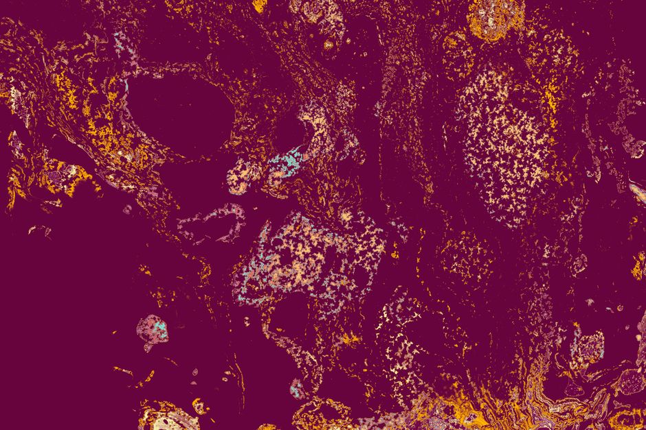
CD31 staining is a laboratory technique that uses a colored marker to highlight a specific protein found on the surface of endothelial cells — the cells that line blood vessels. In diagnosis, pathologists use it to confirm whether a tissue sample contains blood vessels, to identify tumors that arise from blood vessel cells, and to help determine whether a cancer is invading into surrounding tissue.
The test works by attaching a visible dye to an antibody that binds specifically to the CD31 protein. When a pathologist looks at the stained slide under a microscope, the areas that turn brown or red indicate where CD31 is present. This makes it a practical tool for answering specific questions that routine tissue staining cannot.
What Is CD31 Staining And How Is It Used In Diagnosis?
CD31 — also called PECAM-1, short for platelet endothelial cell adhesion molecule-1 — is a protein found on the surface of endothelial cells, platelets, and some white blood cells. It plays a role in how cells stick to each other and how immune cells move through vessel walls.
In pathology, the relevant fact is simpler: CD31 is present on nearly all endothelial cells throughout the body. That makes it a reliable marker for identifying blood vessels in tissue samples.
The staining process itself follows standard immunohistochemistry methods. A thin slice of tissue is placed on a glass slide. The tissue is treated to make the CD31 protein accessible to antibodies. Then a primary antibody that binds to CD31 is applied, followed by a detection system that produces a visible color — typically brown — at the site of binding. A counterstain, usually blue, colors the rest of the tissue for contrast.
What the pathologist sees is straightforward. Any cell or structure that turns brown contains CD31. Blood vessels light up. The pattern and distribution of that staining help answer the diagnostic question at hand.
What Conditions Can CD31 Staining Help Identify?
The most common use of CD31 staining is in tumor pathology. When a pathologist examines a suspicious growth, one key question is whether it originated from blood vessels or from some other tissue type. CD31 staining helps answer that.
Vascular tumors — growths that arise from blood vessel cells — typically show strong CD31 staining because the tumor cells themselves are endothelial in origin. Examples include angiosarcoma, hemangioendothelioma, and benign vascular lesions like hemangiomas. The staining pattern helps distinguish these from tumors that merely have a rich blood supply but are not vascular in origin.
Beyond vascular tumors, CD31 staining helps in several other situations:
- Confirming that a tumor has invaded into blood vessels (vascular invasion), which can affect staging and treatment decisions for some cancers
- Measuring microvessel density in tumors — a research technique that counts how many small blood vessels are present, though its clinical usefulness varies by cancer type
- Identifying endothelial cells in inflammatory conditions where the type of vessel involvement matters
- Distinguishing vascular malformations from other soft tissue lesions
It is worth being clear about what CD31 staining does not do. It does not diagnose cancer by itself. It does not determine whether a tumor is benign or malignant. It answers one specific question — is this structure made of endothelial cells? — and that answer gets combined with other findings to reach a diagnosis.
How Does CD31 Compare To Other Blood Vessel Markers?
CD31 is not the only marker for endothelial cells. Pathologists often use a panel of stains rather than relying on one alone. Each marker has strengths and limitations.
| Marker | What It Stains | Key Limitation |
|---|---|---|
| CD31 | Endothelial cells, platelets, some white blood cells | Can stain plasma cells and some other cells, leading to false positives in certain contexts |
| CD34 | Endothelial cells, some stem cells, certain soft tissue tumors | Less specific for endothelial cells than CD31 in some settings |
| Factor VIII | Endothelial cells | Less sensitive than CD31 — may miss small vessels |
| ERG | Endothelial cells (nuclear staining) | Nuclear pattern can be easier to interpret in some tumors |
| Podoplanin (D2-40) | Lymphatic endothelial cells | Marks lymphatic vessels more than blood vessels |
In practice, a pathologist might order CD31 and CD34 together, or CD31 and ERG, depending on what question needs answering. The choice depends on the tissue type, the suspected diagnosis, and the individual lab’s validated protocols.
One non-obvious point: CD31 can be positive in some cells that are not endothelial. Plasma cells, for example, can show CD31 staining. This is why no single stain is interpreted in isolation. The pathologist looks at the overall pattern, the cell shape, the tissue architecture, and the results of other stains before drawing a conclusion.





When Is CD31 Staining Ordered?
A pathologist orders CD31 staining when the routine examination of a tissue sample raises a question that standard stains cannot answer. It is not a first-line test for most conditions.
Common scenarios include:
- A tumor that appears to contain many blood vessels, raising the possibility of a vascular origin
- A soft tissue mass where the cell type is unclear on routine staining
- A cancer where vascular invasion needs to be assessed for staging purposes
- A skin or mucosal lesion where the pathologist suspects a vascular proliferation
- Evaluation of a liver or bone marrow biopsy where blood vessel patterns are abnormal
The test is performed on tissue that has already been removed — through biopsy, surgical excision, or other sampling procedures. It is not a blood test or an imaging study. The result comes as a pathology report that describes the staining pattern and integrates it with other findings.
What Are The Limitations Of CD31 Staining?
No stain is perfect. CD31 staining has several limitations that are well recognized in pathology practice.
Specificity is one issue. While CD31 is highly expressed on endothelial cells, it can also be found on platelets, macrophages, and some plasma cells. This means a positive result does not automatically mean the stained cells are endothelial.
Sensitivity can vary. In poorly preserved tissue or after certain types of processing, CD31 staining may be weak or absent even when endothelial cells are present. This can lead to false negatives.
Interpretation requires experience. Distinguishing true vascular invasion from artifact, or recognizing when staining is nonspecific, depends on the pathologist’s training and familiarity with the tissue type.
These limitations are not unique to CD31. They apply to immunohistochemistry in general. The strength of the technique comes from using it as part of a panel, combined with careful examination of the tissue architecture and clinical context.
How Should Patients Understand A CD31 Staining Result?
If you see “CD31” on a pathology report, it means the pathologist used this stain as part of the diagnostic workup. The result will be described in the context of the overall findings.
A positive result means the stained cells contain CD31 protein. In most cases, this confirms that the cells are endothelial or that blood vessels are present in the sample. A negative result means the stain did not detect CD31 in the areas examined.
What matters most is how the pathologist interprets the result alongside other information. A positive CD31 stain in a tumor, for example, might support a diagnosis of a vascular tumor — or it might simply show that the tumor has a rich blood supply, which is common in many cancers. The same result can mean different things depending on the context.
Patients who have questions about their pathology report should ask their treating physician to explain what the findings mean for their specific situation. The report is written for clinicians, and the clinical significance depends on the full picture — not on any single stain result.
Frequently Asked Questions
What does a positive CD31 stain mean?
A positive CD31 stain means the tested tissue contains cells that express the CD31 protein, which usually indicates endothelial cells or blood vessels. The clinical meaning depends on what tissue was tested and what other findings are present.
Is CD31 staining the same as a cancer test?
No, CD31 staining is not a cancer test by itself. It is a tool that helps pathologists identify blood vessel cells and can support a cancer diagnosis when combined with other stains and clinical findings.
Can CD31 staining be negative even when blood vessels are present?
Yes, CD31 staining can be falsely negative if the tissue was poorly preserved or if the staining technique did not work optimally. Pathologists often use additional vascular markers to confirm findings when results are unclear.
How long does CD31 staining take?
CD31 staining is typically performed on already-processed tissue and usually takes hours to a day to complete in the lab, depending on the facility’s workflow. The result is then reviewed by a pathologist and included in the final report.
