
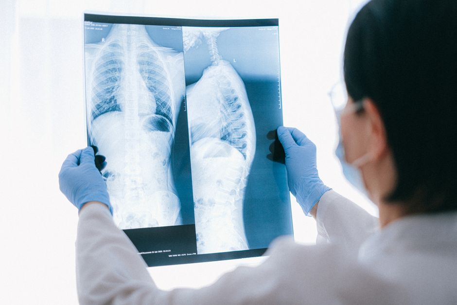
Atelectasis is the partial or complete collapse of a section of lung tissue. When the tiny air sacs called alveoli deflate, that patch of lung can no longer take part in gas exchange. The result is a smaller lung volume and, depending on how much tissue is involved, some degree of low blood oxygen. It is a common finding on chest imaging and a frequent complication after surgery, but it is not a disease on its own — it is a sign of something else going wrong.
The word comes from Greek roots meaning “incomplete expansion.” Doctors use it for both a lung that never fully inflated (as in a newborn) and a lung that was inflated and then collapsed. Most adults who hear the term are in the second group.
What Is Atelectasis Of Lungs?
Atelectasis means airless lung. The alveoli — the grape-like clusters where oxygen crosses into the blood — lose their air and shrivel. Because the affected area still gets blood flow but no air, blood passing through it returns to the heart without picking up oxygen. That mismatch is why atelectasis can lower oxygen levels even when the rest of the lung is healthy.
There are two broad patterns. In obstructive atelectasis, something blocks the airway leading to that region, so the air already inside gets absorbed and the alveoli collapse. In non-obstructive atelectasis, the airway is open but the lung still deflates — because of pressure from outside, because the patient is not breathing deeply, or because surfactant (the substance that keeps alveoli from sticking shut) is missing or reduced.
Size matters. A small patch — sometimes called subsegmental or plate-like atelectasis — is often minor and may cause no symptoms at all. A whole lobe collapsing is a different situation and usually produces noticeable breathing problems.
What Causes Atelectasis?
The causes split along the same obstructive and non-obstructive line, and the list is long because almost anything that interferes with normal lung inflation can produce it.
Airway blockage
When a bronchus is plugged, the air downstream is absorbed into the bloodstream and the alveoli collapse. Common culprits include mucus plugs, especially in people with asthma, cystic fibrosis, or chronic bronchitis. A tumor can block an airway, which is why persistent atelectasis in one spot — particularly in a smoker — needs investigation rather than reassurance. Inhaled foreign objects cause it too, most often in young children. Enlarged lymph nodes can also press on an airway from outside.
Pressure from outside the lung
Air or fluid in the pleural space — the thin gap between lung and chest wall — pushes on the lung and squeezes it shut. This happens with pneumothorax (collapsed lung from air), pleural effusion (fluid), and masses in the chest. A large abdominal swelling or pregnancy can push the diaphragm up and compress the lung bases.
Shallow breathing and immobility
This is the most common cause in hospitalized patients. After surgery, especially chest or abdominal surgery, pain makes people take small, guarded breaths. Lying flat for long periods does the same. The alveoli in the lung bases are small to begin with, and without periodic deep inflation they tend to close off. This is why early walking and deep-breathing exercises are standard after major surgery.
Surfactant problems and other causes
Surfactant reduces surface tension so alveoli stay open. In premature infants, the lungs may not make enough surfactant, producing respiratory distress syndrome and widespread atelectasis. In adults, severe lung injury, prolonged mechanical ventilation, and smoke inhalation can damage surfactant function. Scarring (fibrosis) and certain neuromuscular conditions that weaken the breathing muscles also contribute.
What Are The Symptoms Of Atelectasis?
Small areas of atelectasis often cause no symptoms and are found only on a chest X-ray or CT scan done for another reason. That is common and usually not alarming by itself.
When more lung is involved, symptoms appear:
- Shortness of breath, especially with activity
- A dry cough or one that brings up mucus
- Chest pain, sometimes sharp on breathing
- Fast, shallow breathing
- Low oxygen saturation on a pulse oximeter
- In severe cases, bluish lips or fingertips from low oxygen
Fever can occur with atelectasis, which surprises people. Collapsed lung tissue can trigger a mild inflammatory response that raises temperature. This matters because fever after surgery is sometimes blamed on infection when atelectasis is the actual cause — though infection must still be ruled out, since the two can look alike and can occur together.
How Is Atelectasis Diagnosed?
Imaging is the core of diagnosis. A chest X-ray often shows a collapsed area as increased density — whiter than normal lung — along with signs like a shifted trachea or a raised diaphragm if a whole lobe has collapsed. Chest CT gives a much clearer picture and can reveal the cause, such as a mucus plug or tumor blocking an airway.
Doctors also look at oxygen levels through pulse oximetry or arterial blood gas testing. Lung sounds heard through a stethoscope may be reduced over the affected area. In some cases, a bronchoscopy — passing a thin scope into the airways — is used both to see what is blocking an airway and to clear it.
One useful distinction: atelectasis is a physical finding, not a diagnosis in itself. The imaging shows the collapse; the real question is why it happened. The answer shapes what comes next.
How Is Atelectasis Treated?
Treatment depends entirely on the cause and the size of the collapse. A small patch in a recovering surgical patient often resolves on its own as breathing deepens. A collapsed lobe from a tumor will not improve until the tumor is addressed.
For atelectasis caused by shallow breathing or retained mucus, the main approaches are:
- Deep breathing and coughing — the simplest and most effective measure for post-surgical collapse
- Incentive spirometry — a device that encourages slow, deep breaths
- Early mobilization — getting out of bed and walking
- Chest physiotherapy — positioning and techniques to help clear mucus
- Bronchoscopy — to remove a mucus plug or foreign object blocking an airway
Doctors may also use positive pressure breathing treatments to help reopen closed alveoli. Oxygen is given when blood oxygen is low, though oxygen alone does not reinflate collapsed tissue — it treats the consequence, not the cause.
When atelectasis is caused by a pleural effusion or pneumothorax, draining the fluid or air relieves the pressure and the lung typically re-expands. If a tumor or enlarged lymph node is the cause, treatment targets that underlying problem.
It is worth being clear about what the evidence shows and does not. Deep breathing and early mobilization after surgery are widely used and supported by clinical practice, but the strength of trial evidence varies across specific techniques. Some approaches, like incentive spirometry, are standard in many hospitals even though studies have not consistently shown they outperform simple deep breathing. That does not make them useless — it means the evidence is not as firm as the routine use suggests.
Can Atelectasis Be Prevented?
Much of the prevention advice targets the most common setting: surgery and hospitalization. Because post-operative atelectasis is so common, hospitals build prevention into routine care.
Measures that help include:
- Getting out of bed and walking as soon as it is safe after surgery
- Using an incentive spirometer as instructed
- Controlling pain well enough to breathe deeply — under-treated pain is a major reason people take shallow breaths
- Quitting smoking before planned surgery, which reduces mucus and improves lung function
- Sitting upright rather than lying flat for long periods
For people with conditions that cause thick mucus — cystic fibrosis, for example — regular airway clearance is part of ongoing care and helps prevent collapse. For children, keeping small objects away from reach reduces the risk of inhaling something that blocks an airway.
Prevention is not fully possible. Some atelectasis happens despite good care, especially after major chest or abdominal surgery. It is a common complication, not a sign that something was done wrong.
Is Atelectasis Serious?
Most atelectasis is mild and temporary. A small patch in a hospitalized patient often clears within days as breathing normalizes. It is one of the most frequent findings on post-operative chest imaging and usually resolves without lasting harm.
The picture changes with extent and cause. A large collapse — a whole lobe or an entire lung — can cause significant low oxygen and breathing difficulty, and in rare cases leads to respiratory failure. Atelectasis that persists or keeps returning in the same spot raises concern for a tumor or other blockage and warrants a closer look. In people with existing lung disease, even a modest collapse can tip the balance.
Atelectasis can also set the stage for pneumonia. Collapsed, airless lung tissue is more vulnerable to infection, and the two conditions sometimes develop together. That is one reason doctors take post-operative collapse seriously rather than dismissing it as harmless.
The honest summary: small and short-lived is usually minor; large, persistent, or unexplained is a reason to investigate. The size and the cause decide the seriousness, not the word itself.
Frequently Asked Questions
Is atelectasis the same as a collapsed lung?
Not exactly. “Collapsed lung” usually refers to pneumothorax, where air leaks into the space around the lung and pushes it down. Atelectasis is collapse of lung tissue itself, often from a blocked airway or shallow breathing, and the two have different causes and treatments.
Can atelectasis go away on its own?
Small areas often do, especially after surgery, as breathing deepens and mucus clears. Larger collapses or those caused by a blockage usually need treatment directed at the cause.
Does atelectasis turn into pneumonia?
It can increase the risk. Collapsed, airless lung tissue is more vulnerable to infection, and pneumonia and atelectasis sometimes occur together, which is why persistent collapse is taken seriously.
Is atelectasis cancer?
No, atelectasis is not cancer. But a tumor blocking an airway can cause atelectasis, so collapse that persists in one area — especially in a smoker — should be checked to rule out a blockage.
