
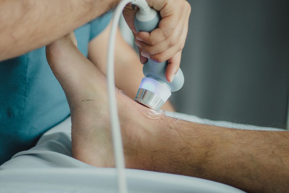
An aorta ultrasound is a painless imaging test that uses sound waves to create pictures of your aorta — the large blood vessel that carries blood from your heart to the rest of your body. It is most often done to look for an abdominal aortic aneurysm, a bulging or weakened section of the aorta that can grow silently for years without causing symptoms. The test takes only a few minutes and gives your doctor a clear view of the aorta’s size and shape.
What Is An Aorta Ultrasound And Why Is It Performed?
An aorta ultrasound — often called an abdominal aortic ultrasound or AAA screening — uses a handheld device called a transducer to send high-frequency sound waves into your abdomen. The sound waves bounce off the aorta and other structures, and a computer turns those echoes into images on a screen.
The main reason doctors order this test is to check for an abdominal aortic aneurysm (AAA). An aneurysm means part of the aorta has stretched and weakened. It can grow slowly over many years. Most people with one feel nothing at all. The danger is that a large aneurysm can rupture, which is a life-threatening emergency.
Finding an aneurysm early is the whole point. When doctors know an aneurysm exists, they can measure it, watch it over time, and plan treatment before it becomes an emergency. Ultrasound is well suited for this because it is quick, uses no radiation, and is inexpensive compared with other imaging methods.
The test can also help evaluate other problems, such as narrowing of the aorta or blockages in the arteries leading to the kidneys and legs. But screening for aneurysm is by far the most common use.
Who Should Get An Aorta Ultrasound?
The strongest risk factor for an abdominal aortic aneurysm is smoking, and the link is well established. Age matters too — aneurysms become more common as people get older. Men develop them more often than women.
Because of this pattern, major medical organizations have recommended a one-time screening ultrasound for certain older men with a history of smoking. The U.S. Preventive Services Task Force has recommended one-time screening for men aged 65 to 75 who have ever smoked. For men in that age range who never smoked, the recommendation has been more selective. For women, routine screening has generally not been recommended unless there is a strong family history or another specific reason.
Screening guidance can be updated as new evidence comes in, so the exact age range and criteria your doctor follows may reflect the most current version. It is reasonable to ask your clinician whether you fall into a group that should be screened.
Other factors that raise risk include:
- A family history of aortic aneurysm
- High blood pressure
- Age — risk rises steadily after about age 60
- Being male
- Having other blood vessel disease, such as narrowed arteries in the legs
One point worth knowing: an aneurysm and a heart attack are not the same problem, even though both involve the circulatory system. Aneurysm screening looks at the wall of the aorta, not at the coronary arteries that supply the heart.
How Do You Prepare For The Test?
Preparation is simple. In most cases you will be asked not to eat or drink for several hours before the exam. This matters because food and gas in the stomach and intestines can block the sound waves and make the aorta harder to see.
Wear loose, comfortable clothing. You will likely be given a gown and asked to lie on your back on an exam table. The technologist will expose your abdomen, from just below the breastbone down to the pelvis.
Tell the technologist or your doctor if you have had abdominal surgery, a stoma, or any open wounds in the area. These do not usually prevent the test, but they may change how it is done.





What Happens During An Aorta Ultrasound?
The exam is quick and does not hurt. A technologist — called a sonographer — applies a clear gel to your abdomen. The gel helps the sound waves travel from the transducer into your body.
The sonographer then presses the transducer gently against your skin and moves it around to get different views. You may feel light pressure, and you may be asked to hold your breath briefly or roll onto your side. The whole study usually takes about 15 to 30 minutes.
You can go back to your normal activities right afterward. There is no recovery period, no sedation, and no radiation exposure.
Sometimes the technologist cannot get a clear picture. Body size, gas, or scar tissue can get in the way. If that happens, your doctor may order a different type of scan, such as a CT scan, to get the information needed.
What Do The Results Mean?
The report will include a measurement of your aorta’s diameter. This number is the key piece of information. The normal size of the abdominal aorta is roughly 2 centimeters across, though it varies with body size and sex.
Doctors generally describe an aneurysm as present when the aorta measures 3 centimeters or larger. Below that, the vessel is considered normal or mildly enlarged. The exact cutoffs and follow-up schedules are clinical decisions your doctor makes based on your measurement, age, sex, and other health factors.
| Aorta Size | General Meaning |
|---|---|
| About 2 cm | Typical normal size |
| Under 3 cm | Normal or mildly enlarged — usually no aneurysm |
| 3 cm or larger | Considered an aneurysm; size and growth rate guide next steps |
If an aneurysm is found, size is not the only thing that matters. How fast it is growing matters too. Smaller aneurysms are often monitored with repeat ultrasounds at intervals set by your doctor. Larger ones may need evaluation for surgical or minimally invasive repair. The decision is made case by case, usually with input from a vascular specialist.
It is worth being clear about one thing: having a small aneurysm is not the same as needing immediate surgery. Many small aneurysms are watched for years and never require an operation.
What Are The Risks And Limitations?
An aorta ultrasound has no known serious risks. It uses sound waves, not radiation, so there is no exposure concern. The gel may feel cool, and the pressure from the transducer may be mildly uncomfortable, but that is the extent of it for most people.
The main limitation is image quality. If the sonographer cannot see the aorta clearly, the test may not give a complete answer. This is not a failure of the test itself — it is a physical limitation of how sound waves travel through the body.
Ultrasound also cannot tell you everything. It shows the size and shape of the aorta well, but other imaging may be needed to plan surgery or to look at certain details. Your doctor will interpret the results in the context of your full health picture.
How Does It Compare With Other Imaging Tests?
Ultrasound is usually the first choice for screening because it is accurate for measuring the abdominal aorta, involves no radiation, and costs less than CT or MRI. For a simple screening question — is there an aneurysm, and how big is it? — ultrasound does the job well.
CT scans give more detailed pictures and are often used when surgery is being planned or when ultrasound results are unclear. They do involve radiation. MRI gives excellent detail without radiation but takes longer, costs more, and is not always easy for people who feel anxious in enclosed spaces.
For routine screening in people without symptoms, ultrasound remains the practical starting point. That is a matter of broad clinical agreement, not a close call.
What Should You Do If Something Is Found?
First, get the actual measurement. Ask your doctor what your aorta measured and how that compares with the threshold for concern. A number gives you something concrete to track.
Second, ask about follow-up. If monitoring is recommended, find out how often and why. Regular follow-up is how small aneurysms are safely watched.
Third, focus on what you can influence. Not smoking is the single most important step for anyone concerned about aortic health. Managing blood pressure and cholesterol also matters, and these are conversations worth having with your clinician.
This article is informational only. It is not a substitute for advice from your own doctor, who knows your history and can interpret your results in context.
Frequently Asked Questions
Does an aorta ultrasound hurt?
No, it does not hurt. You may feel mild pressure from the transducer and the gel may feel cool, but most people find the exam comfortable.
How long does an aorta ultrasound take?
Usually about 15 to 30 minutes. You can return to normal activities right afterward with no recovery time.
Do I need to fast before an aorta ultrasound?
In most cases yes, for several hours before the test. Food and gas can block the sound waves, so your doctor will give you specific instructions.
Can an aorta ultrasound detect a heart problem?
No, it looks at the aorta, not the heart or its arteries. A separate test such as an echocardiogram is used to evaluate the heart.
