
A visual field test measures how much you can see around the edges of your vision while looking straight ahead. It maps the entire area your eye can detect, including your peripheral vision. Doctors use this test to check for blind spots and to monitor conditions that affect your sight over time.
What Is A Visual Field Test and How Does It Work?
A visual field test checks the full span of what your eyes can see. When you stare at a fixed point, you still see objects to the sides, above, and below. That entire range is your visual field.
The test creates a map of your vision. It shows where you see clearly and where you might have gaps or blind spots. These gaps are called scotomas. Everyone has one natural blind spot where the optic nerve connects to the retina, but other gaps can signal eye disease.
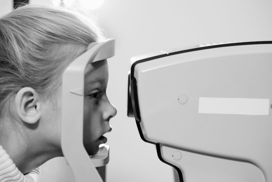
During the test, you look into a machine called a perimeter. You focus on a central target while lights flash in different areas. Each time you see a flash, you press a button. A computer records which lights you saw and which you missed.
The test takes about 15 to 30 minutes per eye. It requires concentration, but there is no physical discomfort. Your eye does not need to be touched.
Why Would a Doctor Order a Visual Field Test?
Doctors order visual field tests to diagnose and monitor several eye conditions. The most common reason is glaucoma, a disease that damages the optic nerve. Glaucoma often starts by causing small blind spots in peripheral vision. These spots grow slowly, and many people do not notice them until significant vision is already lost.
The test also helps detect and track other conditions, including:
- Optic nerve damage from injury or disease
- Retinal conditions such as retinitis pigmentosa
- Neurological problems like strokes or brain tumors that affect vision pathways
- Multiple sclerosis, which can cause optic neuritis
- Side effects from certain medications, particularly some anti-epilepsy drugs
Visual field tests are also used before and after eye surgery. Cataract surgery can change vision, and doctors may want a baseline before operating. For people with glaucoma, the test is usually repeated at regular intervals to see if the disease is stable or progressing.
What Are the Different Types of Visual Field Tests?
Several methods exist for measuring the visual field. The most common is automated perimetry. You sit at a machine, place your chin on a rest, and respond to flashing lights. The machine runs a preset pattern and produces a detailed printout of your results.
Another type is confrontation visual field testing. This is a quick screening method done in a regular exam room. The doctor sits across from you and asks you to count fingers or identify moving objects in your peripheral vision. It is less precise than automated testing but can catch large visual field defects.
Kinetic perimetry uses moving targets instead of flashing lights. A technician moves a light slowly inward from the edges of your vision. You press a button when you first see it. This method is useful for mapping the outer boundaries of your visual field.
Amsler grid testing is a simple home-based test for the central visual field. You look at a grid of lines and note any areas that appear wavy, blurred, or missing. This test is often used for people with macular degeneration.




What Do the Results Mean?
Your results come as a printed map with shaded areas showing your vision sensitivity. Darker areas indicate reduced sensitivity. The computer compares your results to an average person of the same age.
A normal visual field extends about 60 degrees upward, 75 degrees downward, 100 degrees outward, and 60 degrees inward from the center. These numbers vary slightly by individual, and the test accounts for normal variation.
Blind spots on the map can indicate different problems depending on their location and shape. Small, localized gaps near the center may suggest early glaucoma. Large gaps on one side might point to a stroke or brain lesion. The pattern of vision loss gives your doctor important clues about the underlying cause.
Your doctor will interpret the results in context. A single abnormal test does not always mean you have a disease. People can perform poorly on a test due to fatigue, anxiety, or misunderstanding the instructions. Doctors often repeat the test to confirm findings.
How Should You Prepare for a Visual Field Test?
Preparation is simple. Get a good night of sleep before the test. Fatigue can slow your reaction time and affect your results.
If you wear glasses or contact lenses, bring them to your appointment. You will need your best corrected vision for the test. If you normally use reading glasses, you may need them for the test as well.
Avoid drinking alcohol for at least a few hours before the test. Alcohol can affect your reaction time and visual perception. Some medications can also influence results, so tell your doctor about everything you take.
You may want to arrange a ride home if your eyes will be dilated. Dilation drops make your vision blurry and your eyes sensitive to light for several hours. Not every visual field test requires dilation, but some do.
Are There Risks or Side Effects?
Visual field testing is safe. It involves no radiation and no contact with your eye. The only requirement is that you sit still and respond to visual stimuli.
The main downside is fatigue. Staring at a central point for 20 minutes while watching for flashes is mentally tiring. Your eyes may feel strained afterward, but this passes quickly.
If you have eye drops for dilation, you may experience blurred vision and light sensitivity for a few hours. Wearing sunglasses after the test can help with the brightness.
There are no known long-term risks from visual field testing. It is safe to repeat as often as your doctor recommends.
How Often Should You Have a Visual Field Test?
The frequency depends on your condition. People with stable glaucoma often have the test once or twice a year. Those with more advanced disease may need it every few months.
People without eye disease may never need a visual field test. It is not part of a routine eye exam for everyone. Your doctor will recommend it if you have symptoms or risk factors that warrant checking.
If you notice changes in your vision between appointments, contact your eye doctor. Sudden vision loss, new blind spots, or a feeling like a curtain is coming over your eye are urgent warning signs that need immediate attention.
Frequently Asked Questions
Does a visual field test hurt?
No, the test is completely painless. You simply look into a machine and press a button when you see lights.
How long does a visual field test take?
The test typically takes 15 to 30 minutes per eye. The exact time depends on the type of test and how carefully you respond.
Can I drive after a visual field test?
You can usually drive if your eyes were not dilated. If dilation drops were used, your vision may be blurry and you should not drive until it clears.
What happens if my visual field test is abnormal?
An abnormal result does not automatically mean you have a serious condition. Your doctor will likely repeat the test and may order additional imaging or exams to determine the cause.
