
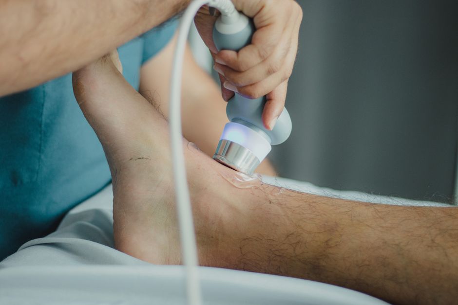
A renal ultrasound is a painless imaging test that uses sound waves to create pictures of your kidneys and the blood vessels and tissues around them. It does not use radiation. The test usually takes about 30 minutes, and most people need little to no preparation. Results are typically read by a radiologist and sent to your doctor, often within a few days.
What Is A Renal Ultrasound Procedure Prep Results?
A renal ultrasound — sometimes called a kidney ultrasound — is a noninvasive scan that shows the size, shape, and position of your kidneys. It can also detect fluid buildup, blockages, stones, cysts, and masses. A handheld device called a transducer is moved across your belly or lower back. It sends out high-frequency sound waves that bounce off your organs and create live images on a screen.
No needles. No radiation. No recovery time. You can usually drive yourself home and go back to your normal day right after.
The test is different from a CT scan or an MRI. A CT scan uses X-rays. An MRI uses magnets and radio waves. Both can image the kidneys, but each has different strengths and tradeoffs. A renal ultrasound is often the first imaging choice because it is safe, quick, and does not expose you to radiation. It is also the preferred option during pregnancy when other imaging may not be appropriate.
What the procedure actually involves
You lie on an exam table, usually on your back. A technologist applies a clear gel to your abdomen or flank. The gel helps the sound waves travel from the transducer into your body. The technologist presses the transducer against your skin and moves it around to capture views of both kidneys and your bladder.
You may be asked to take a deep breath and hold it for a few seconds. That moves your kidneys into a better position for imaging. The whole exam is generally painless. If you have a full bladder, the pressure from the transducer may feel mildly uncomfortable, but it passes quickly.
After the scan, you wipe off the gel and you are done. There is no downtime.
How Do You Prepare for a Renal Ultrasound?
Preparation is minimal for most people. Many patients need no preparation at all. Your doctor’s office will give you specific instructions, and those instructions take priority over any general guidance.
The most common preparation is drinking water before the exam so your bladder is full. A full bladder pushes certain structures into a clearer view and lets the technologist see your bladder itself. You may be asked to drink a set amount of water about an hour before your appointment and not urinate until after the scan.
Some facilities ask you to avoid eating for several hours beforehand, especially if your doctor also wants images of nearby organs. Others do not restrict food at all. This varies by clinic and by what your doctor is looking for.
Tell the technologist if you:
- Are pregnant or think you might be
- Have a urinary catheter or a drainage tube
- Had recent kidney surgery or a biopsy
- Have open sores or a rash on your abdomen
Wear loose, comfortable clothing. You will likely lift your shirt or change into a gown. Leave jewelry at home if it might get in the way, though the exam does not usually require removing metal the way an MRI does.
One thing worth knowing: a renal ultrasound does not require contrast dye. That matters if you have kidney disease or a contrast allergy, because it removes a concern that comes with some CT scans. It also means you do not need to fast for contrast or arrange a ride home afterward.
What Can a Renal Ultrasound Detect?
A renal ultrasound can identify several structural problems in and around the kidneys. It is especially good at showing blockages, fluid collections, and differences in kidney size or shape.
Common findings include:
- Kidney stones — especially stones in the kidney itself, though very small stones or those in the ureter can be harder to see
- Hydronephrosis — swelling of a kidney caused by urine backing up
- Cysts — fluid-filled sacs that are often harmless but sometimes need monitoring
- Kidney size and symmetry — one kidney much smaller than the other can signal a chronic problem
- Masses or tumors — solid growths that may need further imaging
- Fluid around the kidney — which can point to injury or other conditions
- Bladder problems — such as incomplete emptying
The test also helps monitor known conditions over time. If you have chronic kidney disease, polycystic kidney disease, or a history of stones, your doctor may order repeat ultrasounds to track changes.
What it does not do well is measure how well your kidneys are filtering blood. That is a job for blood tests like creatinine and estimated glomerular filtration rate. Imaging shows structure. Blood tests show function. Doctors often use both together.
What Do Renal Ultrasound Results Mean?
A radiologist reviews the images and writes a report for your doctor. The report describes what was seen and usually includes an impression — a short summary of the key findings.
Results generally fall into three rough categories:
- Normal — both kidneys appear normal in size, shape, and position, with no blockages or abnormal fluid
- Abnormal but not urgent — findings like simple cysts or mild differences in kidney size that need monitoring but not immediate action
- Findings that need follow-up — blockages, solid masses, or significant fluid that may require more imaging, a referral, or a procedure
Your doctor will explain what the findings mean for you specifically. A result that sounds alarming in the report may be minor in context, and a “normal” ultrasound does not rule out every possible kidney problem. Some conditions, especially those affecting how the kidney works rather than how it looks, will not show up on an ultrasound at all.
Timing varies. Some clinics give results the same day. Others take a few days. Ask when you can expect to hear back so you are not left waiting without a plan.
What Are the Risks and Limitations?
A renal ultrasound has no known risks when performed by a trained technologist. It uses sound waves, not radiation, and there is no evidence of harm from diagnostic ultrasound at standard settings. This is why it is considered safe during pregnancy.
The limitations are about what it can and cannot see, not about safety.
Ultrasound waves do not pass well through bone or air. That means the test can struggle to image certain parts of the urinary tract, particularly the ureters — the tubes that carry urine from the kidneys to the bladder. A stone lodged in a ureter may not show up clearly. Gas in the bowel can also block the view.
Body size matters too. More tissue between the transducer and the kidney can reduce image quality. In those cases, a doctor may recommend CT or MRI instead.
Ultrasound also cannot tell you how well your kidneys are working. It cannot measure filtration. And it cannot always distinguish between a harmless cyst and a solid mass with certainty — sometimes follow-up imaging is needed to be sure.
None of this makes the test less useful. It just means your doctor may combine it with other tests to get the full picture.
When Is a Renal Ultrasound Ordered?
Doctors order a renal ultrasound for many reasons. It is often a first step because it is safe, fast, and gives useful information without radiation.
Common reasons include:
- Blood in the urine
- Pain in the flank or lower back
- Frequent urinary tract infections
- Poor kidney function on blood tests
- High blood pressure that is hard to control
- A known or suspected kidney stone
- Monitoring a known kidney condition
- Evaluating an injury to the abdomen or back
- Checking the kidneys before certain surgeries or procedures
It is also used to guide procedures. During a kidney biopsy, for example, the doctor may use ultrasound to see exactly where the needle is going.
In children, ultrasound is often the first imaging choice for urinary problems because it avoids radiation. The same is true for pregnant women, where it can safely check for kidney swelling or stones without exposing the fetus to X-rays.
How Does a Renal Ultrasound Compare to Other Kidney Tests?
Different imaging tests answer different questions. The right choice depends on what your doctor needs to know.
| Test | Best For | Radiation | Contrast Dye |
|---|---|---|---|
| Ultrasound | Structure, fluid, stones, size | None | No |
| CT scan | Stones, masses, detailed anatomy | Yes | Often |
| MRI | Soft tissue, complex masses | None | Sometimes |
| Blood tests | Kidney function (filtration) | None | No |
CT is generally better at finding small stones, especially in the ureter. MRI gives the most detail for soft tissue and complex masses. But both have tradeoffs — CT involves radiation and often contrast dye, and MRI is slower and more expensive.
Ultrasound is often the starting point. If it answers the question, no further imaging may be needed. If it does not, your doctor can move to another test with a clearer idea of what to look for.
Blood tests and urine tests are not alternatives to ultrasound — they are complements. They tell you about function and infection. Ultrasound tells you about structure.
Does a Renal Ultrasound Require Contrast Dye?
No. A standard renal ultrasound does not use contrast dye. That is one of its main advantages.
Contrast dye used in CT scans and some MRI scans can be a concern for people with reduced kidney function or a history of allergic reactions. Ultrasound avoids that issue entirely. It also means no fasting for contrast, no IV line, and no need to arrange a ride home.
In some specialized cases, a doctor may use an ultrasound contrast agent — tiny gas-filled bubbles injected into a vein. This is not the same as CT or MRI contrast, and it is not routine. It is used in specific situations and only at centers that offer it.
For the standard kidney ultrasound most people receive, there is no dye, no needle, and no radiation.
Frequently Asked Questions
Do I need to fast for a renal ultrasound?
Most people do not need to fast, but some clinics ask you to avoid food for several hours beforehand. Follow the specific instructions from your doctor’s office, since they take priority over general guidance.
How long does a renal ultrasound take?
The exam usually takes about 30 minutes. You can typically return to normal activities immediately afterward with no recovery time.
Can a renal ultrasound detect kidney cancer?
It can detect a solid mass in the kidney, which may prompt further imaging to determine whether it is cancer. Ultrasound alone often cannot confirm whether a mass is cancerous, so CT or MRI is usually needed for a clearer answer.
Is a renal ultrasound painful?
The exam itself is not painful. You may feel mild pressure from the transducer, especially if your bladder is full, but this passes quickly.
