
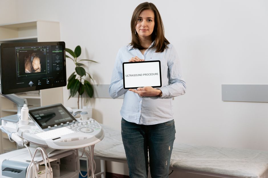
A hysterosonogram is an ultrasound procedure that looks inside your uterus to find the cause of abnormal bleeding or other uterine problems. A thin tube is placed through the cervix and a small amount of sterile salt water is used to gently expand the uterine cavity. The fluid acts as a window, allowing the ultrasound probe to produce clear images of the inside of the uterine wall. This is why it is also called a saline infusion sonogram or simply a “water ultrasound.”
How Does A Hysterosonogram Work?
Ultrasound uses sound waves to create pictures. A standard pelvic ultrasound can see the outside shape of the uterus, but it cannot see the inside lining well. The lining is collapsed and pressed together, like two wet pieces of paper stuck flat.
The hysterosonogram solves that problem. A physician inserts a small, soft catheter into the cervix. Then a sterile saline solution flows slowly into the uterine cavity. The fluid pushes the walls of the uterus apart, creating a clear space. The ultrasound probe on your abdomen then sends sound waves through that fluid to create a detailed image of the inner lining.
This technique gives the doctor a direct view of the endometrial lining. It shows the shape of the cavity and any growths attached to the wall. It is a diagnostic test, not a treatment. It helps the doctor understand what is happening inside the uterus so they can recommend the right next step.
Why Would A Doctor Order A Hysterosonogram?
Doctors order this test to investigate specific symptoms or abnormal findings. The most common reason is abnormal uterine bleeding. This includes heavy periods, bleeding between periods, or bleeding after menopause.
The procedure is also used to evaluate an abnormal finding on a regular ultrasound. If a standard ultrasound shows a possible polyp, fibroid, or thickened lining, a hysterosonogram provides a closer look. It can confirm whether a growth exists, where it is attached, and how large it is.
It is also used for women who have had recurrent miscarriages. The test can reveal structural problems in the uterus, such as a septum or scar tissue, that might interfere with pregnancy. Infertility evaluations sometimes include this test for the same reason.
Doctors also use it before certain fertility treatments. Knowing the exact shape of the uterine cavity helps the physician plan the best approach. It is a functional test that gives real answers about the internal environment of the uterus.
How Do You Prepare For The Procedure?
Preparation is simple but important. The test is usually scheduled during the first half of your menstrual cycle, typically between days 5 and 10. That timing avoids pregnancy and ensures the lining is thin enough to see clearly.
You should tell your doctor if there is any chance you might be pregnant. The procedure is not performed on a pregnant woman. You should also mention any known allergies, especially to latex or iodine, and list all medications you take.
Your doctor may recommend taking an over-the-counter pain reliever about an hour before the appointment. Ibuprofen or acetaminophen can reduce cramping during and after the test. Do not take anything without asking your doctor first, especially if you have medical conditions or take other medications.
You will be asked to empty your bladder before the procedure. A full bladder pushes the uterus into a position that makes it harder to see. You will change into a hospital gown and lie on an exam table with your feet in stirrups, similar to a Pap smear.





What Happens During The Procedure?
The entire appointment usually takes 20 to 30 minutes. The active part of the procedure typically lasts less than 10 minutes. You are awake the whole time.
The doctor first performs a standard transvaginal ultrasound to get a baseline view of the uterus and ovaries. Then a speculum is inserted into the vagina to hold it open, just like during a Pap smear. The cervix is cleaned with an antiseptic solution.
A thin, flexible catheter is then gently passed through the cervix and into the uterine cavity. This is the most uncomfortable part of the procedure. Most women describe it as mild to moderate cramping, similar to menstrual cramps. The speculum is removed, and the ultrasound probe is reinserted into the vagina.
The doctor slowly injects the saline solution through the catheter. You will feel fluid moving and possibly more cramping as the uterus expands. The doctor watches the screen and takes images while the fluid fills the cavity. The fluid drains out through the catheter and the vagina after the test is complete.
What Do The Results Show?
The images reveal the internal structure of the uterine cavity. A normal result shows a smooth, triangular-shaped cavity with no growths or obstructions. The lining appears even and uniform in thickness.
Abnormal findings are common and specific. Polyps appear as smooth, round growths attached to the lining. Fibroids appear as solid masses that may push into the cavity. Scar tissue, called adhesions, looks like bands or webs crossing the cavity. A uterine septum appears as a band of tissue dividing the cavity partially or completely.
These findings matter because they have different implications. Polyps and fibroids are usually benign but can cause bleeding and fertility problems. Scar tissue can interfere with implantation. A septum is a structural difference present from birth that may increase miscarriage risk.
Your doctor will explain the findings and recommend next steps. In many cases, a follow-up procedure can remove the growth. The hysterosonogram itself does not treat anything. It is a mapping tool that guides the treatment plan.
What Are The Risks And Side Effects?
The procedure is safe when performed by a trained clinician. Serious complications are rare. The most common side effect is cramping during and shortly after the test. Light spotting for a day or two is normal and expected.
Infection is uncommon but possible. The risk is low because sterile equipment is used. Call your doctor if you develop fever, heavy bleeding, severe pelvic pain, or foul-smelling discharge after the procedure. These could be signs of infection.
There is a small risk of the catheter perforating the uterine wall. This is extremely rare and more likely in women with a thin uterus or significant scar tissue. Most perforations heal on their own without treatment.
Vasovagal reactions can occur. This is a drop in heart rate and blood pressure triggered by cervical stimulation. It causes lightheadedness or fainting. It is temporary and resolves quickly once the procedure stops.
How Does It Compare To Other Uterine Tests?
Several tests look at the uterus, and they are not interchangeable. A standard pelvic ultrasound sees the outside shape but not the inside lining clearly. A hysterosonogram adds fluid to see the inner cavity directly.
A hysteroscopy is a different procedure. It uses a small camera inserted through the cervix to look directly inside the uterus. It provides a live, direct view and can also be used to perform surgery at the same time. It is more invasive and usually requires anesthesia.
An endometrial biopsy samples the lining tissue for laboratory testing. It checks for abnormal cells or cancer but does not show the shape of the cavity. It answers a different question than a hysterosonogram.
Saline infusion sonography is sometimes confused with sonohysterography. They are the same test with different names. The choice of test depends on your symptoms and what your doctor needs to know.
Frequently Asked Questions
Is a hysterosonogram painful?
Most women feel mild to moderate cramping during the procedure, similar to menstrual cramps. The discomfort usually lasts only a few minutes and eases quickly after the fluid is drained.
How long does a hysterosonogram take?
The entire appointment takes about 20 to 30 minutes. The actual fluid infusion and imaging portion typically takes less than 10 minutes.
Can I go back to work after a hysterosonogram?
Yes, most women can resume normal activities immediately after the procedure. Light spotting and mild cramping are common for a day, so many women prefer to take it easy for the rest of the day.
Will a hysterosonogram affect my ability to get pregnant?
No, the procedure does not harm fertility. In fact, it is often used during fertility evaluations to check the uterine cavity for problems that could interfere with pregnancy.
