
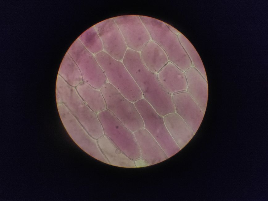
Columnar cell change is a benign finding in breast tissue. It means that some of the cells lining the milk ducts have taken on a taller, more columnar shape than usual. It is not cancer, and by itself it does not raise your risk of breast cancer.
Most women who have this finding never know it. It turns up on a breast biopsy that was done for another reason. The word “change” can sound alarming, but in this case it describes a common and harmless variation in how cells look under a microscope.
What Does Columnar Cell Change In The Breast Mean?
It means the cells lining the terminal ductal lobular units — the small milk-producing structures deep in the breast — have changed shape. Instead of the flat or cuboidal appearance of normal duct lining, they look taller and more tightly packed, like columns.
This is a microscopic finding. It cannot be seen on a mammogram, felt during an exam, or detected by ultrasound. A pathologist only sees it after examining a tissue sample under a microscope.
Columnar cell change is common. It is found in a meaningful share of breast biopsies performed for other reasons, particularly in women in their 40s and 50s. Many pathologists consider it part of the normal spectrum of breast tissue changes that can happen with age and hormonal shifts.
The key point: columnar cell change alone is not considered a risk factor for breast cancer. It does not require treatment. It does not need to be removed. In most cases, it simply gets noted in a pathology report and that is the end of it.
How Is Columnar Cell Change Different From Columnar Cell Hyperplasia?
This distinction matters, and it is where confusion often starts. Columnar cell change and columnar cell hyperplasia are related but not identical.
In columnar cell change, the cells are taller than usual but there are still only one or two layers of them. The architecture stays simple. In columnar cell hyperplasia, there are more layers of cells — typically more than two — and the cells may cluster or form small tufts. The term “hyperplasia” means increased cell growth.
Both are benign. Both are part of a group of findings sometimes called columnar cell lesions. The difference is mainly about how many cell layers are present and how complex the pattern looks under the microscope.
Some research suggests that columnar cell hyperplasia, particularly when it appears alongside certain other findings like atypia, may be associated with a slightly increased risk of breast cancer over time. But the evidence here is not uniform, and many studies have not found a meaningful increase in risk when columnar cell hyperplasia appears on its own without atypia.
This is an area where pathologists and oncologists do not fully agree. Some clinicians recommend more frequent screening for women with columnar cell hyperplasia. Others do not. If you receive this diagnosis, the most useful thing you can do is ask your doctor whether any atypia was seen in the same sample — because that is the finding that changes the conversation.
What Causes Columnar Cell Change?
The exact cause is not known. What is known is that these changes are linked to hormone exposure, particularly estrogen. They are rarely seen in women before menopause and become more common with age.
Columnar cell change is sometimes found alongside other benign breast conditions like fibrocystic changes, which also involve hormone-related tissue shifts. It is not caused by anything you did or did not do. It is not linked to diet, stress, deodorant use, or wearing tight bras.
One clarification worth making: columnar cell change is not the same as atypical ductal hyperplasia or lobular carcinoma in situ. Those are different findings with different implications. If your pathology report mentions columnar cell change but does not mention atypia, those other diagnoses are not present.





Does Columnar Cell Change Raise Your Risk Of Breast Cancer?
On its own, no. Columnar cell change without atypia is not considered a risk factor for breast cancer by major pathology and oncology organizations.
The picture changes if atypia is present. Atypia means the cells look abnormal in ways that are not cancer but are not fully normal either. When columnar cell change appears together with flat epithelial atypia — a specific type of atypia sometimes found in these lesions — some studies suggest a modest increase in breast cancer risk. Other studies have not confirmed this.
The evidence is genuinely mixed. This is not a situation where you should assume the worst, and it is also not one where you should ignore the finding if atypia is mentioned.
If your report says “columnar cell change without atypia,” your risk is not increased by this finding. If it says “columnar cell change with flat epithelial atypia” or “with atypia,” ask your doctor what that means for your screening schedule. Some clinicians recommend yearly mammograms in that situation. Others recommend the same schedule as for women without this finding. There is no single standard.
What Happens After A Diagnosis Of Columnar Cell Change?
In most cases, nothing beyond routine care. Columnar cell change does not need surgery, medication, or any specific follow-up beyond what you would normally do for breast health.
If the finding was made on a core needle biopsy, your doctor may recommend a surgical excisional biopsy to make sure nothing else is nearby. This is not because columnar cell change is dangerous. It is because core needle biopsies sample only a small amount of tissue, and there is a small chance that a more significant finding could be present in the surrounding area. Whether this step is needed depends on what else was seen in the sample and your overall risk profile.
If the finding was made on an excisional biopsy — where the whole area was removed — no further action is typically needed for the columnar cell change itself.
Routine breast cancer screening recommendations still apply. For most women aged 40 to 74 at average risk, that means mammograms every one to two years, though exact schedules vary by organization and personal history. Columnar cell change does not change those recommendations unless atypia is also present.
What Should You Ask Your Doctor?
The pathology report is the most important document here. Ask for a copy if you do not have one. Then ask these questions:
- Does the report mention atypia, flat epithelial atypia, or any other atypical finding?
- Was this found on a core biopsy or an excisional biopsy?
- Do you recommend any additional imaging or follow-up because of this finding?
- Does this change my screening schedule?
If the answer to the first question is no, and the finding was on an excisional biopsy, you likely do not need to think about it again. If the answer is yes, or if the biopsy was a core needle sample, a more detailed conversation is reasonable.
It also helps to know that columnar cell change is not rare. Pathologists see it regularly. It is one of many benign findings that show up in breast tissue as women age. The word “change” does not mean “pre-cancer.” It means the cells look different from the textbook picture of normal — and in this case, that difference is not dangerous.
Is There Anything You Should Do Differently?
No. There is no diet, supplement, exercise routine, or lifestyle change that has been shown to reverse or prevent columnar cell change. No clinical guidelines recommend any specific intervention for this finding alone.
General breast health recommendations still apply — maintaining a healthy weight, limiting alcohol, and staying current with screening. But these are general recommendations for all women, not specific to columnar cell change.
If you are postmenopausal and taking hormone therapy, that is a separate conversation to have with your doctor about your overall risk profile. Columnar cell change itself is not a reason to stop or change hormone therapy.
Frequently Asked Questions
Is columnar cell change in the breast cancer?
No. Columnar cell change is a benign finding, not cancer, and it does not become cancer on its own.
Does columnar cell change increase breast cancer risk?
Columnar cell change without atypia does not increase breast cancer risk. If atypia is also present, some studies suggest a modest increase, but the evidence is mixed.
Should columnar cell change be removed?
No. Columnar cell change does not need to be removed. Your doctor may recommend a follow-up biopsy if the sample was small, but this is to check for other findings, not because the columnar cell change itself is dangerous.
What is the difference between columnar cell change and columnar cell hyperplasia?
Columnar cell change has one or two layers of taller cells, while columnar cell hyperplasia has more than two layers and may show small cell clusters. Both are benign.
