
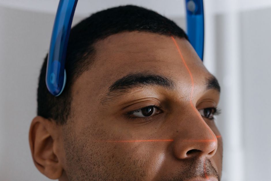
An orbital CT scan shows detailed cross-sectional images of the bony eye socket, the eyeball, the optic nerve, and the muscles and fat that surround the eye. Doctors use it most often to look for fractures after facial trauma, to find infections or tumors in or behind the eye, and to check for foreign objects such as metal or glass. It is fast, widely available, and especially good at showing bone.
What Does An Orbital Ct Scan Show?
An orbital CT scan produces thin, layered images of the orbit — the cone-shaped cavity of bone that holds the eye. A computer stacks those slices into views a radiologist can scroll through from top to bottom, side to side, and front to back.
What it can reveal includes:
- Bones: The orbital rim, the thin floor and medial walls, and the optic canal. These thin bones break easily in facial trauma, and CT shows fractures clearly.
- The eyeball: Its shape, position, and the density of the fluid inside. Blood or a mass inside the eye can change that density.
- The optic nerve: Its size and path from the back of the eye toward the brain.
- Extraocular muscles: The muscles that move the eye, including whether they are enlarged or thickened.
- Orbital fat and blood vessels: The soft tissue cushioning the eye.
- The sinuses and brain edge: The ethmoid and maxillary sinuses sit right against the orbit, so problems there can affect the eye.
CT is built around X-rays, so it distinguishes bone and dense material very well. It is less detailed than MRI for soft tissue like nerves and small tumors. That trade-off matters when choosing which scan to order.
When Is An Orbital CT Scan Ordered?
Trauma is the most common reason. A blow to the face, a fall, or a car accident can fracture the orbit, and doctors need to know whether bone has shifted, whether a muscle is trapped, or whether the eye itself is injured.
Other common reasons include:
- Suspected infection spreading from the sinuses into the orbit, which can threaten vision
- Bulging or protruding eye (proptosis) with no clear cause
- Double vision or restricted eye movement
- Vision loss that needs urgent evaluation
- Suspected foreign body, such as metal or glass near the eye
- A known or suspected tumor in or around the orbit
CT is often the first choice in an emergency because it is quick and available around the clock. MRI usually gives better detail for soft tissue and may be ordered afterward when the question is about nerves, tumors, or inflammation rather than bone.
What Can A CT Scan Miss Or Not Show Well?
CT is not the best test for everything. It has real limits, and knowing them helps you understand why a doctor might order a second scan.
Soft tissue detail is the main limitation. Small tumors, early inflammation of the optic nerve, and some vascular problems may be hard to see or characterize on CT. MRI generally shows these better.
Radiation is another consideration. CT uses ionizing radiation, and the dose to the eye lens is a known concern. The lens is one of the more radiation-sensitive tissues in the body. For a single scan the risk is generally considered small, but repeated scans add up. This is one reason doctors avoid CT when the question could be answered another way.
CT also cannot always tell what a mass is made of. It can show that something is there and where it sits, but distinguishing a benign growth from a malignant one often requires MRI, a biopsy, or both. A normal-looking CT does not rule out every problem.





What Does The Procedure Involve?
An orbital CT is short and usually painless. You lie still on a table that slides into a ring-shaped scanner. The scan itself often takes only a few minutes.
In many cases a contrast dye is used. It is given through an IV and can make certain tissues and blood vessels easier to see. If you have kidney problems, a history of reactions to contrast, or are pregnant, tell the staff beforehand — contrast and radiation both need extra thought in those situations.
You may be asked to remove metal objects like earrings, hairpins, or glasses. Metal can create streaks on the images that obscure nearby detail. For a suspected metallic foreign body near the eye, staff follow specific safety steps, since a strong magnetic field is not involved in CT but other precautions still apply.
Afterward you can usually go back to normal activities right away. If contrast was used, drinking water helps your body clear it.
How Do Doctors Use The Results?
The images go to a radiologist, who reviews them and writes a report. Your doctor then combines that report with your symptoms, your exam, and any other tests.
What happens next depends on what the scan shows. A fracture may need surgery or may heal on its own. An infection may need antibiotics or drainage. A mass may need MRI, a biopsy, or monitoring over time. A scan that looks normal in someone with ongoing symptoms may still prompt further testing, because imaging is one piece of the picture, not the whole answer.
One point worth understanding: an orbital CT is a tool for finding structural problems. It does not measure how well you see, and it does not replace a careful eye exam. Two people can have similar scans and very different symptoms.
Orbital CT Compared With Other Eye Imaging
Different scans answer different questions. This table summarizes the general strengths of each.
| Test | Best For | Main Limitation |
|---|---|---|
| CT | Bone, fractures, foreign bodies, acute trauma | Less soft-tissue detail; uses radiation |
| MRI | Nerves, tumors, inflammation, soft tissue | Slower; not ideal for metal foreign bodies |
| Ultrasound | Structures inside the eye; no radiation | Cannot see through bone |
| X-ray | Quick check for some fractures | Limited detail; misses many injuries |
The choice is not about which test is “best” overall. It is about which one answers the specific question at hand with the least risk and the most useful detail.
Is An Orbital CT Scan Safe?
For most people, a single orbital CT is considered low risk, and the information it provides often outweighs that risk. The main safety considerations are radiation and contrast dye.
Radiation exposure from one scan is small, but it is not zero, and the eye lens is relatively sensitive to it. This is why doctors try to avoid unnecessary repeat scans and why they weigh alternatives when they can. If you are pregnant or think you might be, tell your doctor before the scan so the team can decide whether it is needed or whether another test would work better.
Contrast dye can cause allergic reactions in some people and can affect kidney function in others. Most reactions are mild, but serious reactions are possible. Sharing your medical history, allergies, and medications in advance helps the team plan safely.
The evidence does not support the idea that CT is risk-free, nor does it support avoiding a needed scan out of fear. The reasonable position is that CT is a valuable, well-established tool whose use should be matched to a clear clinical question.
Frequently Asked Questions
What does an orbital CT scan show that a regular eye exam cannot?
It shows the bone, the optic nerve, the eye muscles, and the tissue behind the eye in cross-section, which a physical exam cannot see. This makes it useful for fractures, foreign bodies, and problems deep in the socket.
Is an orbital CT scan the same as a brain CT?
No, they cover different areas and answer different questions, though they are sometimes done together. An orbital CT focuses on the eye socket, while a brain CT looks at the brain itself.
Does an orbital CT scan use radiation?
Yes, CT uses ionizing radiation, and the eye lens is a relatively radiation-sensitive tissue. A single scan is generally considered low risk, but repeated scans should be justified by a clear medical need.
Can an orbital CT scan detect a tumor behind the eye?
It can often show that a mass is present and where it sits, but it may not be able to tell what the mass is. MRI or a biopsy is frequently needed to characterize it further.
