
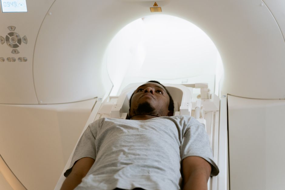
A hypoattenuating area on a CT scan is a region that appears darker than the tissue around it. It means that area absorbs less X-ray energy than neighboring tissue, so fewer X-rays are blocked and the spot shows up gray or near-black instead of bright. The term describes an appearance, not a diagnosis — it tells you how a piece of tissue behaves on the scan, and your doctor interprets what that appearance most likely represents.
That distinction matters. “Hypoattenuating” is a descriptive word radiologists use, and it can point to something harmless like a cyst filled with fluid or something that needs follow-up like a tumor. The scan alone rarely delivers the final answer.
What Does A Hypoattenuating Area On A Ct Scan Mean?
It means that specific patch of tissue is less dense — less physically packed — than the tissue around it. Dense structures like bone block a lot of X-rays and appear white (hyperattenuating). Air blocks almost none and appears black. A hypoattenuating area sits somewhere on that scale toward the darker end.
Radiologists measure this density in Hounsfield units (HU), a standardized scale. Water is defined as 0 HU. Air is about −1000 HU. Bone runs into the hundreds and higher. Fat sits roughly between −50 and −100 HU. Knowing where a lesion falls on this scale helps a radiologist narrow down what it is made of.
Here is the part many people miss: a single CT image is not enough to characterize a hypoattenuating area. Radiologists look at how the area behaves before and after intravenous contrast dye is given. Some lesions light up (enhance) with contrast because they have a blood supply. Others stay dark because they are fluid-filled and have no vessels. That enhancement pattern often matters more than the initial darkness.
What Usually Causes a Hypoattenuating Area?
The most common explanations are fluid-filled structures: simple cysts, fluid collections, or areas of swelling. Fluid is less dense than solid tissue, so it reads as hypoattenuating. Many of these are benign and require nothing more than a note in the report.
Other causes depend heavily on which organ is being scanned. In the liver, a hypoattenuating spot could be a cyst, a fatty patch, an abscess, or a tumor. In the brain, it could reflect an area of old injury, swelling, or reduced blood flow. In the kidney, it might be a cyst or a collection of urine. The same word covers all of these because the word describes density, not disease.
Fat is another frequent cause. Fatty tissue is hypoattenuating compared with normal organ tissue, which is why a fatty liver looks darker than a healthy one on CT. This is a good example of how the finding can reflect a common, manageable condition rather than anything alarming.
Does Hypoattenuating Mean Cancer?
No. Most hypoattenuating areas are not cancer. Many are simple cysts or benign growths that never cause a problem. The word itself carries no cancer diagnosis.
That said, some cancers do appear hypoattenuating, which is exactly why the finding gets investigated rather than dismissed. A radiologist weighs several features together: the shape and borders of the area, whether it enhances with contrast, its exact density, its location, and how it compares with any earlier scans. A smooth, round, fluid-density spot with thin walls is far more likely to be a cyst. An irregular area with thick walls that enhances with contrast raises more concern.
This is also why your doctor may order more imaging — an ultrasound, an MRI, or a repeat CT — or recommend a biopsy. Each test adds information the first scan could not provide. A CT finding on its own is a clue, not a verdict.




How Do Radiologists Tell the Difference Between Causes?
They combine density measurements with contrast behavior and anatomy. The Hounsfield unit value gives a first clue about what the tissue is made of. Contrast enhancement shows whether it has a blood supply and how that supply behaves. Location and shape narrow the list further.
Here is a rough guide to how density readings point in different directions. These are general reference ranges, not diagnostic thresholds — the interpretation always depends on the organ and the clinical picture.
- Near −1000 HU: air or gas
- Roughly −50 to −100 HU: fat
- Around 0 HU: simple fluid, as in a water-density cyst
- Higher values: soft tissue, with the exact number varying by organ and by whether contrast was used
A lesion that stays at fluid density and does not enhance is often a simple cyst. One that enhances strongly after contrast suggests a solid structure with blood vessels. One with mixed density may be more complex. Radiologists also compare the area to the surrounding normal tissue, because “hypoattenuating” is always relative to something.
What Happens After a Hypoattenuating Area Is Found?
What happens next depends entirely on context: where the area is, how it looks, your symptoms, and your medical history. There is no single standard next step for every hypoattenuating finding.
Sometimes the radiologist can confidently call it benign — a classic simple cyst, for example — and no further action is needed. Other times the report recommends follow-up imaging to confirm the area is stable over time. Occasionally a biopsy is needed to know for certain. In urgent situations, such as a suspected abscess or a sign of reduced blood flow to an organ, the finding may prompt immediate treatment.
Your own doctor puts the scan together with your symptoms and history. A hypoattenuating spot in someone with fever and pain means something different than the same spot found by chance in someone feeling well. The imaging finding is one piece of a larger picture.
What Should You Ask Your Doctor About the Finding?
Ask what the area most likely represents and whether it needs follow-up. Request the specific language from the radiology report, because “hypoattenuating” alone does not tell you the size, location, or whether it enhanced with contrast.
A few questions tend to be useful:
- Where exactly is the area, and how large is it?
- Did it enhance with contrast, and what does that suggest?
- Does the report recommend follow-up imaging, and if so, when?
- Does this finding connect to any symptoms I am having?
- Is a biopsy or another test needed, or can we watch and wait?
It also helps to know whether you have prior scans for comparison. A hypoattenuating area that has been stable for years is usually reassuring, while a new or growing one gets more attention. If you have had imaging elsewhere, make sure your current doctor can access those images.
The Bottom Line
A hypoattenuating area is a description of how tissue looks on a CT scan — darker and less dense than its surroundings. It is not a diagnosis on its own. The meaning comes from the full picture: the density, the contrast behavior, the location, your symptoms, and any prior scans.
Most such findings turn out to be benign, but some require follow-up or further testing. The right move is not to panic or to ignore it, but to get a clear explanation from the doctor who ordered the scan and to follow through on any recommended next steps.
Frequently Asked Questions
Is a hypoattenuating area on a CT scan serious?
It depends on what the area represents, and many turn out to be harmless cysts or fatty patches. Some do require follow-up testing, so the finding should always be interpreted by your doctor rather than judged from the word alone.
Does hypoattenuating mean fluid?
Often it does, because simple fluid reads as darker and less dense than solid tissue on CT. Fat and some solid lesions can also appear hypoattenuating, which is why contrast behavior and density measurements help tell them apart.
Can a hypoattenuating area be cancer?
Some cancers do appear hypoattenuating, but most hypoattenuating areas are not cancer. Radiologists use shape, borders, density, and contrast enhancement to judge the likelihood and decide whether more testing is needed.
Will I need a biopsy for a hypoattenuating area?
Not always — many findings are diagnosed from the scan alone or with follow-up imaging. A biopsy is usually reserved for areas that remain uncertain or look concerning after other tests.
