
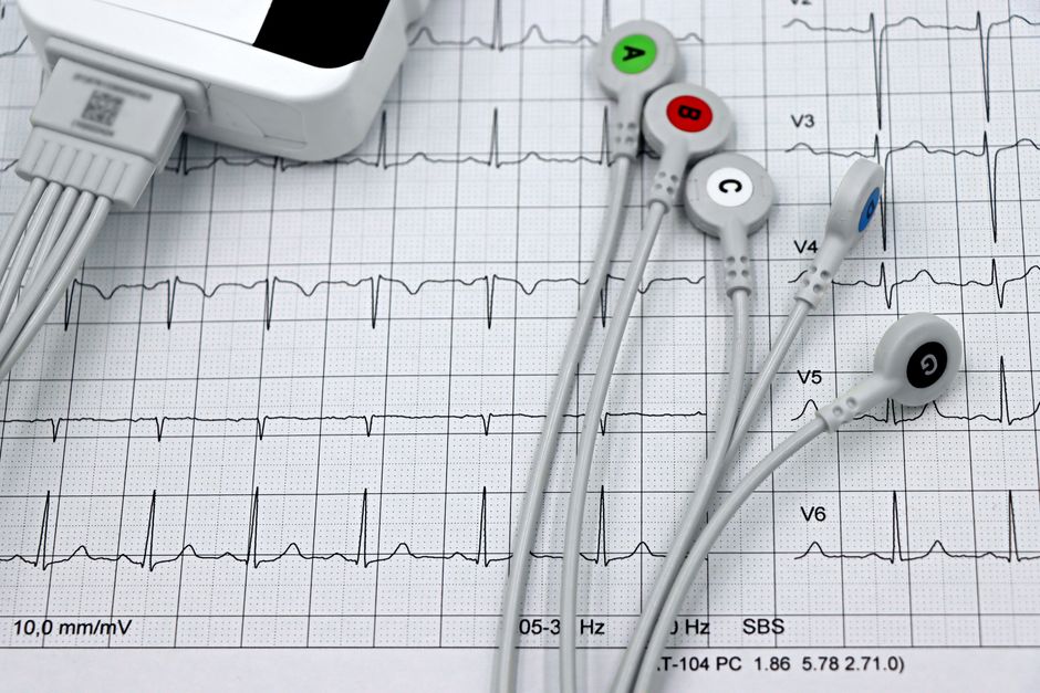
The precordial leads on an ECG — the six chest electrodes labeled V1 through V6 — show the heart’s electrical activity from a horizontal plane. They provide a detailed view of the front and side walls of the heart, specifically the septum, the anterior wall of the left ventricle, and the lateral wall. These leads are essential for locating heart muscle damage during a heart attack and for identifying certain rhythm and conduction problems.
What Are the Precordial Leads on an ECG?
The precordial leads are the six electrodes placed directly on the chest during a standard 12-lead electrocardiogram. They are labeled V1 through V6. Each lead sits at a specific spot on the chest wall. Together they capture electrical signals moving through the heart muscle from different angles.
Limb leads — the other six leads in a 12-lead ECG — look at the heart from vertical and diagonal planes. Precordial leads provide the horizontal or transverse view. This matters because some areas of the heart are best seen from the chest wall, not from the arms or legs.
Standard placement is well established. V1 sits at the fourth intercostal space just to the right of the sternum. V2 sits at the fourth intercostal space just to the left of the sternum. V4 goes at the fifth intercostal space on the midclavicular line. V3 sits directly between V2 and V4. V5 goes at the anterior axillary line at the same horizontal level as V4. V6 sits at the midaxillary line at that same level.
Correct placement matters. Even small shifts can change the tracing and lead to misdiagnosis.
What Do the Precordial Leads (V1-V6) Show About Heart Activity?
Each precordial lead sees a specific region of the heart. V1 and V2 face the interventricular septum — the wall between the left and right ventricles. V3 and V4 face the anterior wall of the left ventricle. V5 and V6 face the lateral wall of the left ventricle.
On a normal ECG the precordial leads show a predictable pattern. The R wave — the first upward deflection — starts small in V1 and gradually gets taller moving toward V6. This is called normal R-wave progression. The S wave — the first downward deflection after the R wave — does the opposite. It is deepest in V1 and shallowest in V6.
Doctors look at this pattern closely. Loss of normal R-wave progression can indicate an old heart attack, left ventricular hypertrophy, or a conduction problem. A very tall R wave in V1 with a deep S wave in V6 can suggest right ventricular hypertrophy. The opposite pattern — deep S in V1 with tall R in V6 — can suggest left ventricular hypertrophy.
The ST segment and T wave on precordial leads are also critical. ST elevation in specific leads points to acute heart muscle injury in that region. ST depression can indicate ischemia or reciprocal changes from another area.
How Do Precordial Leads Help Diagnose Heart Attacks?
Precordial leads are essential for localizing a heart attack. Different coronary arteries supply different parts of the heart. When a blockage cuts blood flow the affected muscle shows characteristic changes on the ECG — usually ST elevation, then Q waves over time.
Changes in V1 and V2 suggest a septal heart attack. This usually involves the left anterior descending artery. Changes in V3 and V4 suggest an anterior heart attack — also typically the left anterior descending artery. Changes in V5 and V6 suggest a lateral heart attack, which can involve the left circumflex artery or a branch of the left anterior descending artery.
When multiple precordial leads show changes together the pattern helps determine the extent of damage. Extensive anterior ST elevation across V1 through V4 signals a large heart attack affecting much of the front wall of the heart. This type carries higher risk and requires aggressive treatment.
Importantly, some heart attacks affect the back or inferior wall of the heart. These may not show obvious ST elevation on the standard precordial leads. In those cases the posterior leads — V7, V8, and V9 — placed on the back can be added to complete the picture. This is not part of a standard 12-lead ECG but is used when posterior infarction is suspected.
Research consistently shows that the location and extent of ST elevation on precordial leads correlates with the size of the heart attack and patient outcomes. This is why the 12-lead ECG — including precordial leads — is performed within minutes of arrival at an emergency room for anyone with chest pain.





What Is the Difference Between Precordial and Limb Leads?
Limb leads and precordial leads look at the heart from different angles. Limb leads use electrodes on the arms and legs and capture the heart’s electrical activity in the frontal plane. They divide into standard limb leads — I, II, III — and augmented limb leads — aVR, aVL, aVF.
Precordial leads capture activity in the horizontal plane. This gives a cross-sectional view of the heart from front to back and side to side. Think of the limb leads as seeing the heart from above and below. Think of the precordial leads as seeing it from the front of the chest toward the back.
Each set provides information the other cannot. Limb leads are better for seeing the inferior wall of the heart — leads II, III, and aVF — and the high lateral wall — lead I and aVL. Limb leads also provide the axis of the heart, which helps diagnose conduction problems and chamber enlargement.
Precordial leads are better for the anterior wall, septum, and low lateral wall. Together the 12 leads give a three-dimensional picture. No single lead tells the full story.
A standard ECG always includes both sets. The 12-lead ECG is called “12-lead” because it has 10 physical electrodes — four on the limbs and six on the chest — but produces 12 distinct electrical views.
What Does an Abnormal Precordial Lead Reading Mean?
Many things can make precordial leads look abnormal. The meaning depends on which leads are affected and what the change looks like.
ST elevation in contiguous precordial leads — meaning two or more leads next to each other — is the classic sign of acute heart muscle injury. This is a medical emergency. ST depression in precordial leads can mean ischemia or can be a reciprocal sign of a heart attack on the opposite wall — for example ST depression in V1 and V2 with ST elevation in the inferior leads.
Q waves are deep downward deflections that appear after a heart attack has damaged the heart muscle permanently. A Q wave in V1 or V2 that is deeper than normal can indicate a prior septal or anterior heart attack. Q waves do not always mean permanent damage, but in most cases they represent scar tissue.
Poor R-wave progression — where the R wave does not get taller normally from V1 to V4 — can signal an old anterior heart attack. It can also occur with left ventricular hypertrophy or with lung disease that changes the position of the heart in the chest.
A very tall R wave in V1 with a deep S wave in V6 is a pattern of right ventricular hypertrophy. This can happen with conditions that strain the right side of the heart, such as pulmonary hypertension or severe lung disease. The opposite pattern — deep S in V1, tall R in V6 — suggests left ventricular hypertrophy from high blood pressure or aortic valve disease.
T wave inversions in precordial leads can mean many things. Some are normal variants — particularly in young adults. Others indicate ischemia, electrolyte disturbances, or even a brain injury called a subarachnoid hemorrhage. The clinical context always matters.
No single abnormal finding on precordial leads confirms a diagnosis by itself. The ECG is one piece of information. Doctors interpret it alongside symptoms, physical exam, blood tests for cardiac enzymes, and sometimes imaging studies.
Frequently Asked Questions
Why are there six precordial leads instead of just one?
Six leads are needed because different chest positions see different regions of the heart. No single chest electrode can adequately see the septum, anterior wall, and lateral wall all at once.
Can the precordial leads detect a heart attack that already happened?
Yes. Pathological Q waves on precordial leads often indicate a prior heart attack in the septal, anterior, or lateral region. The presence and location of Q waves help doctors determine when and where damage occurred.
Do precordial leads show the right ventricle of the heart?
Standard precordial leads do not directly view the right ventricle. Right-sided precordial leads placed on the right side of the chest are needed for that. These are used when right ventricular involvement is suspected.
What happens if a precordial lead is placed in the wrong spot?
Misplacement changes the tracing and can produce false ST elevations or other abnormal patterns that mimic a heart attack. Consistent and correct placement is essential for accurate interpretation.
