
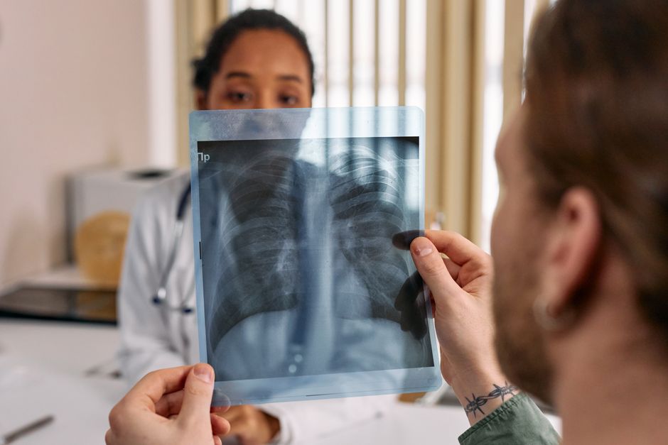
A collapsed lung happens when air, blood, or another substance gets between the lung and the chest wall, or when something blocks the airways so a section of lung deflates. The medical term for the most common type is pneumothorax. It can strike a tall, thin young man with no warning, follow a rib fracture, or develop as a complication of lung disease. The cause determines how dangerous it is and how fast it needs treatment.
What Causes a Lung to Collapse?
The short answer is that a lung collapses when the pressure balance around it changes, or when airflow into it gets blocked.
Your lungs sit inside a sealed space called the pleural cavity. A thin membrane lines the inside of your chest wall and wraps around each lung. Between those two layers is a tiny amount of fluid that lets them slide smoothly as you breathe. Normally the pressure in that space is slightly negative compared to the air pressure inside your lungs. That negative pressure is what keeps your lungs pulled open.
If air leaks into that space, the pressure equalizes or turns positive. The lung gets pushed inward and deflates, like a balloon squeezed from the outside. This is a pneumothorax.
There is a second way a lung can collapse that has nothing to do with the pleural space. If something blocks a bronchial tube, the air trapped beyond the blockage gets absorbed into the bloodstream over time and that section of lung shrinks. Doctors call this atelectasis. It is common after surgery, with mucus plugging, or with a tumor pressing on an airway.
These two mechanisms produce different problems and different treatments. A pneumothorax is usually sudden and often painful. Atelectasis tends to develop quietly and shows up on imaging.
What Are the Different Types of Pneumothorax?
Doctors group pneumothorax into categories based on what caused it and whether the lung is still leaking air.
- Primary spontaneous pneumothorax — happens without an obvious trigger in someone with no known lung disease. It is most common in tall, thin men between their late teens and early 30s. Small blebs, which are weak spots on the lung surface, rupture and leak air.
- Secondary spontaneous pneumothorax — happens in people who already have lung disease. Chronic obstructive pulmonary disease (COPD) is a common cause. So are cystic fibrosis, tuberculosis, and lung cancer.
- Traumatic pneumothorax — follows a chest injury. A broken rib can puncture the lung. A stab wound or gunshot wound can open the chest wall.
- Iatrogenic pneumothorax — caused by a medical procedure. Needle biopsies, central line placement, and mechanical ventilation can all introduce air into the pleural space.
- Tension pneumothorax — a one-way leak that lets air in but not out. Pressure builds with every breath and can push the heart and major blood vessels aside. This is a medical emergency.
The distinction matters because a primary spontaneous collapse in a healthy young person is often manageable with observation or a simple drainage tube. The same size collapse in someone with severe COPD can be life-threatening.
What Causes Atelectasis (Blocked Airway Collapse)?
Atelectasis develops when an airway gets blocked and the air beyond it is gradually absorbed. This is different from a pneumothorax and is far more common in hospital settings.
The most frequent cause is mucus. After surgery, especially chest or abdominal surgery, breathing tends to be shallow and coughs are weak. Mucus collects, plugs a small airway, and the lung tissue downstream deflates. This is why hospitals push patients to take deep breaths and use incentive spirometers after operations.
Other causes include:
- A tumor growing into or pressing on a bronchus
- An inhaled object, more common in children
- Enlarged lymph nodes from infection or cancer
- Prolonged bed rest or immobility
- Severe lung scarring that distorts the airways
Atelectasis usually causes no pain. It may show up as low oxygen levels, a mild cough, or a shadow on a chest X-ray. Large areas of atelectasis can reduce oxygen exchange enough to cause shortness of breath.





Who Is Most at Risk?
Risk factors differ by type, and some are well established while others are less clear.
For primary spontaneous pneumothorax, the strongest known risk factors are being male, being tall and thin, and smoking. Smoking increases risk substantially — some research suggests the risk in smokers is many times higher than in nonsmokers, though the exact multiple varies across studies. A family history also raises risk, which points to a genetic component in some cases.
For secondary pneumothorax, the risk comes from the underlying lung disease. People with COPD who have emphysema-like changes in the lung tissue are at particular risk because the lung surface is weaker.
For atelectasis, the main risk factors are recent surgery, prolonged bed rest, shallow breathing from pain or sedatives, and any condition that weakens the cough reflex.
Age matters differently depending on the type. Primary spontaneous pneumothorax peaks in young adulthood. Secondary pneumothorax becomes more common with age as lung disease accumulates.
What Does a Collapsed Lung Feel Like?
The classic symptom is sudden sharp chest pain on one side, often described as a stabbing sensation, followed by shortness of breath. The pain may radiate to the shoulder or back. It often gets worse with deep breathing or coughing.
Not everyone gets the textbook picture. Some people, especially those with a small primary spontaneous pneumothorax, feel only mild discomfort or a vague sense that something is off. Others have no pain at all and notice only that they cannot catch their breath.
Signs that suggest a more serious situation include:
- Rapid heart rate
- Low oxygen levels
- Blue tint to lips or fingertips
- Severe shortness of breath at rest
- Feeling faint or confused
A tension pneumothorax is a different level of emergency. Blood pressure drops, the neck veins may bulge, and one side of the chest may not move with breathing. This requires immediate decompression — a needle or tube inserted to release the trapped air. Waiting for imaging is not an option in that situation.
How Is a Collapsed Lung Treated?
Treatment depends on the size, the cause, and how much the person is struggling to breathe.
A small primary spontaneous pneumothorax in a stable person may be watched with repeat X-rays, because the body can reabsorb the air over time. This is common practice, though which collapses need a tube versus observation is an area where guidelines have shifted and continue to evolve.
Larger collapses, or any collapse causing significant breathlessness, usually need a chest tube. The tube is placed between the ribs into the pleural space and connected to a drainage system. Air escapes, the lung re-expands, and the tube stays in until the leak seals.
For recurrent collapses or after a second episode, doctors may recommend a procedure to prevent future ones. Options include pleurodesis, where the two pleural layers are deliberately irritated or stuck together so the space closes, and surgical removal of blebs. These are well-established approaches for preventing recurrence, though the best timing and method vary by patient.
Atelectasis is treated differently. The goal is to open the blocked airway. Deep breathing exercises, coughing, chest physical therapy, and sometimes bronchoscopy to clear a mucus plug are standard. If a tumor is the cause, treating the tumor is the priority.
When Should You Seek Emergency Care?
Sudden chest pain with shortness of breath needs immediate medical attention. Do not wait to see if it passes.
Call 911 or go to an emergency room if you have:
- Sudden sharp chest pain, especially on one side
- Shortness of breath that is new or getting worse
- Rapid heartbeat with chest discomfort
- Fainting or near-fainting
- Blue lips or fingertips
These symptoms can also signal a heart attack or pulmonary embolism, which are also emergencies. The cause cannot be sorted out at home. Imaging and an exam are needed.
One point worth clarifying: a collapsed lung is not the same as a punctured lung, though people often use the terms interchangeably. A punctured lung usually means the lung tissue itself has been torn, which may or may not cause a pneumothorax. A collapsed lung refers to the deflation. The distinction matters for treatment.
Frequently Asked Questions
Can a collapsed lung heal on its own?
A small pneumothorax in a stable person can sometimes reabsorb without a procedure, and doctors may monitor it with repeat imaging. Larger collapses or ones causing significant breathing problems usually need a chest tube or other intervention.
What is the most common cause of a collapsed lung?
Primary spontaneous pneumothorax, which happens when a small weak spot on the lung surface ruptures without any obvious trigger, is the most common type in healthy young adults. In hospital settings, atelectasis from mucus plugging after surgery is more common.
Does smoking cause a collapsed lung?
Smoking is a well-established risk factor for primary spontaneous pneumothorax, and the risk increases with how much a person smokes. Quitting reduces that risk over time.
How do you know if your lung has collapsed?
Sudden sharp chest pain on one side with shortness of breath is the classic sign, but some people feel only mild discomfort or nothing at all. Only imaging can confirm it.
