
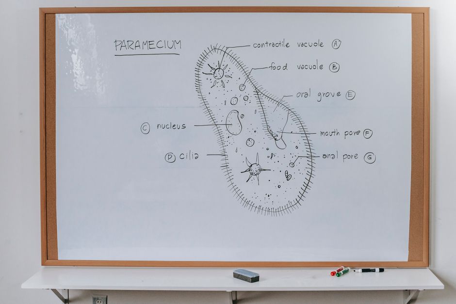
A cilia microscope lets you see tiny hair-like structures called cilia. These are thin extensions that stick out from the surface of many cells in the body. Under a light microscope you can see their movement and general shape. To see their internal structure clearly, you need an electron microscope. That difference matters, because what you can observe depends heavily on which instrument you are using.
Cilia are found in places you might not expect. They line your airways, help move fluid through the fallopian tubes, and sit inside the inner ear helping you sense balance. Some single-celled organisms swim using cilia. Each of these can be viewed under the right microscope, but the level of detail changes with the equipment.
What Can You See With A Cilia Microscope?
With a standard light microscope, you can see cilia as fine, moving fringes along the edge of a cell or tissue sample. They look like tiny hairs beating in waves. At magnifications of around 400x to 1000x, their general shape and motion become visible.
What you cannot see with a light microscope is the internal structure inside each cilium. That requires an electron microscope, which uses beams of electrons instead of light to create images at far higher resolution. With electron microscopy, scientists can see the arrangement of microtubules — the protein tubes that form the core of a cilium.
This internal core, called the axoneme, has a well-established structure. It typically consists of nine pairs of microtubules arranged in a ring, with two single microtubules in the center. This “9+2” pattern is one of the most consistent findings in cell biology. It appears in cilia across many species.
So the answer depends on your instrument:
- Light microscope: overall shape, location, and beating motion of cilia
- Electron microscope: internal microtubule arrangement, membrane details, and structural defects
- Fluorescence microscope: specific proteins within cilia, using labeled markers
What Do Cilia Look Like Under a Light Microscope?
Under a light microscope, cilia appear as a dense brush border of fine projections along one surface of a cell. They are usually too small to see individually at low power. At higher magnification, you can make out individual strands, though they remain at the edge of what light microscopy can resolve.
A single cilium is roughly 0.2 to 0.3 micrometers in diameter. That is close to the limit of resolution for a standard light microscope, which is about 0.2 micrometers. This means individual cilia are barely resolvable. You see them best as a group.
Living cilia are often observed in motion. A tissue sample from the airway lining, for example, can show cilia beating in coordinated waves. This movement is what helps clear mucus and trapped particles from the lungs. Watching that motion under a microscope is one of the clearest demonstrations of cilia function.
One practical point: cilia are fragile. Sample preparation for microscopy can damage or flatten them, which is why careful handling matters in any laboratory setting that studies them.
What Can Electron Microscopy Reveal About Cilia?
Electron microscopy reveals the detailed internal architecture of cilia. The 9+2 microtubule arrangement becomes clearly visible in cross-section. This level of detail is not achievable with light microscopy.
This matters clinically. Some inherited conditions involve defects in cilia structure or function. These are called ciliopathies. In primary ciliary dyskinesia, for example, cilia do not beat normally, which can lead to chronic respiratory infections and other problems. Electron microscopy has historically been used to examine cilia structure in patients being evaluated for such conditions.
That said, structural examination alone does not always confirm or rule out a ciliary disorder. Some genetic defects cause cilia to malfunction without obvious structural changes visible even under electron microscopy. Genetic testing has become an important part of evaluation for these conditions. The role of electron microscopy in diagnosis has shifted somewhat as genetic methods have improved.
Where in the Body Are Cilia Found?
Cilia are widespread in the human body. They serve different functions depending on location.
- Respiratory tract: cilia line the airways and move mucus upward to clear debris and pathogens
- Fallopian tubes: cilia help move the egg toward the uterus
- Inner ear: specialized cilia (stereocilia) are involved in hearing and balance
- Brain ventricles: cilia help circulate cerebrospinal fluid
- Kidney tubules: a single primary cilium on many kidney cells senses fluid flow
Not all cilia are the same. Motile cilia beat in coordinated waves. Primary cilia are non-motile and act more like sensors, detecting signals from the environment around the cell. Under a microscope, the two types can look different in both structure and behavior.
What Can You See in Single-Celled Organisms With Cilia?
Some single-celled organisms, like Paramecium, are covered in cilia that they use to swim and to sweep food toward their mouth-like opening. These are large enough to be seen with a basic light microscope, and their cilia are often visible in motion.
Paramecium is a common subject in school biology classes for exactly this reason. The organism is relatively large, and its cilia beat fast enough to be observed in real time. You can watch the organism move and see the coordinated beating that propels it.
This is one of the easiest ways to see cilia directly without advanced equipment. A drop of pond water under a light microscope can reveal these organisms and their cilia in action.
What Are the Limitations of Cilia Microscopy?
Microscopy has real limits when it comes to cilia. Light microscopes cannot resolve internal structure. Electron microscopes provide that detail but require fixed, processed samples, so you cannot watch living cilia in motion with them.
Sample preparation also affects what you see. Chemical fixation, dehydration, and sectioning can alter cilia shape or damage delicate structures. Results depend on how the sample was handled.
There is also the question of interpretation. Identifying a structural abnormality in cilia requires trained expertise. Not every variation seen under a microscope indicates disease. Normal biological variation exists, and distinguishing it from a true defect takes experience.
The evidence for using cilia microscopy as a diagnostic tool is strongest for specific inherited conditions, and even there it is often used alongside genetic testing rather than alone. For most general health questions, cilia microscopy is a research and specialist tool, not a routine test.
Does Cilia Microscopy Have a Role in Medical Diagnosis?
Cilia microscopy has a defined but limited role in medicine. It is used in specialist settings, particularly for evaluating suspected primary ciliary dyskinesia and related disorders. In these cases, examining cilia structure and beat pattern can provide useful information.
However, no single microscopy finding is definitive on its own. Diagnosis of ciliary disorders typically combines clinical history, microscopy, and genetic testing. The relative weight given to each has changed over time as genetic methods have advanced.
For most people, cilia microscopy is not part of routine medical care. It is a laboratory and research tool. The conditions it helps investigate are uncommon.
Can you see cilia with a regular light microscope?
Yes, but only their general shape and movement. Individual cilia are near the resolution limit of light microscopes, so internal structure is not visible.
What is the 9+2 structure of cilia?
It is the arrangement of microtubules inside most motile cilia, with nine pairs in a ring around two central microtubules. This pattern is visible only with electron microscopy.
Are cilia and stereocilia the same thing?
No. Stereocilia in the inner ear are structurally different from true cilia despite the similar name. They are supported by actin filaments rather than microtubules.
Can cilia defects cause health problems?
Yes. Inherited defects in cilia structure or function can cause conditions called ciliopathies, which may affect the lungs, kidneys, and other organs.
Frequently Asked Questions
Can you see cilia with a regular light microscope?
Yes, but only their general shape and movement. Individual cilia are near the resolution limit of light microscopes, so internal structure is not visible.
What is the 9+2 structure of cilia?
It is the arrangement of microtubules inside most motile cilia, with nine pairs in a ring around two central microtubules. This pattern is visible only with electron microscopy.
Are cilia and stereocilia the same thing?
No. Stereocilia in the inner ear are structurally different from true cilia despite the similar name. They are supported by actin filaments rather than microtubules.
Can cilia defects cause health problems?
Yes. Inherited defects in cilia structure or function can cause conditions called ciliopathies, which may affect the lungs, kidneys, and other organs.