
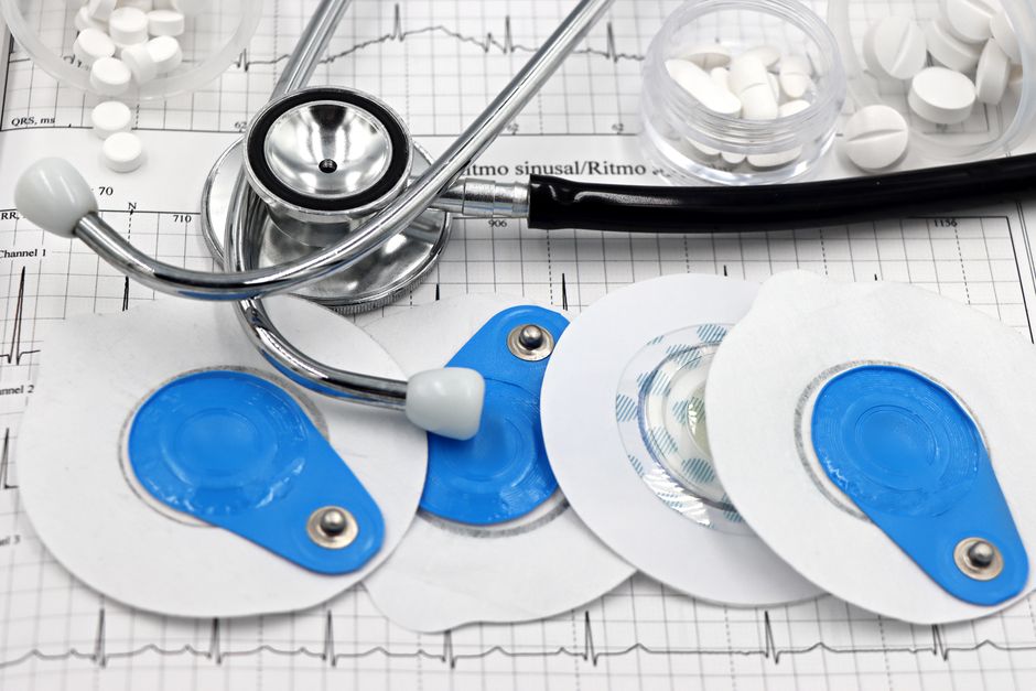
A cardiac stress test makes your heart work harder while a monitor records how it responds. There are several types. The main ones are the exercise treadmill test, the exercise stress test with imaging (either echocardiography or nuclear imaging), the pharmacological stress test for people who cannot exercise, and stress testing with cardiac MRI or CT. Your doctor picks the type based on your symptoms, your ability to exercise, and what question they need answered.
What Are The Different Types Of Cardiac Stress Tests?
Cardiac stress tests fall into two broad groups: tests that use exercise to stress the heart, and tests that use medication to simulate exercise. Within each group, some tests only record the heart’s electrical activity, while others add imaging to show blood flow or wall motion.
The core types are:
- Exercise treadmill test — also called a standard stress ECG. You walk or run on a treadmill while electrodes record your heart rhythm.
- Exercise stress echocardiography — a treadmill or bike test combined with ultrasound images of the heart taken before and immediately after exercise.
- Exercise nuclear stress test — exercise combined with a small amount of radioactive tracer that shows blood flow to the heart muscle.
- Pharmacological stress test — medication is used to increase heart rate or dilate coronary arteries when a person cannot exercise.
- Stress cardiac MRI — magnetic resonance imaging during pharmacological stress to assess blood flow and heart muscle.
- Stress CT perfusion — CT imaging with pharmacological stress to evaluate blood flow, still used mainly in research and select centers.
Each type answers a slightly different question. A standard treadmill test primarily looks at the heart’s electrical response to exertion. Imaging tests look at blood flow or how the heart muscle moves. Choosing between them is a clinical decision, not a matter of one being universally better.
What Happens During a Standard Exercise Treadmill Test?
A standard exercise treadmill test is the simplest form of cardiac stress testing. You walk on a treadmill while a technician and physician monitor your heart rhythm, blood pressure, and symptoms.
The protocol usually follows a standardized sequence. Speed and incline increase every few minutes in stages. The goal is to bring your heart rate to a target based on your age, or until you develop symptoms that require stopping. The test typically takes 15 to 30 minutes including preparation and recovery.
Electrodes placed on your chest record a 12-lead electrocardiogram throughout. The physician watches for changes in the ST segment, which can indicate reduced blood flow to part of the heart muscle. Blood pressure is checked at intervals. You report any chest pain, shortness of breath, dizziness, or leg fatigue.
The test is stopped if you reach your target heart rate, develop significant symptoms, or show concerning ECG changes. A normal result means no electrical evidence of ischemia during the level of exertion you achieved. It does not rule out coronary artery disease entirely, especially if you did not reach an adequate workload.
This test is most useful in people with a moderate likelihood of coronary artery disease and no conditions that make the ECG hard to interpret. It is less reliable in people with certain baseline ECG abnormalities, such as left bundle branch block, or those taking digoxin.
How Does Stress Echocardiography Differ From a Standard Test?
Stress echocardiography adds ultrasound imaging to the stress test. The key difference is that it shows how the heart muscle physically moves, not just its electrical signals.
Images are captured before exercise and within about a minute after peak exercise. The heart is imaged in multiple views. A physician compares the resting and stress images side by side. Areas of muscle that do not contract normally under stress may indicate reduced blood flow.
This approach improves accuracy compared with a standard treadmill test alone, particularly in women and in people whose baseline ECG is difficult to interpret. It also provides information about valve function and heart muscle thickness.
One practical limitation: the images must be obtained quickly after exercise stops, because the heart returns to its resting state within a couple of minutes. Some centers use a supine bike instead of a treadmill to allow imaging during peak exercise, which avoids that time pressure.
The test requires adequate image quality. In some people, body habitus or lung disease makes the heart difficult to visualize clearly.




What Is a Nuclear Stress Test?
A nuclear stress test uses a small amount of radioactive tracer to show blood flow to the heart muscle. It is one of the most established imaging methods for detecting reduced coronary blood flow.
Tracer is injected when the heart is at peak stress and again at rest. A special camera detects the tracer and produces images of the heart muscle. Areas that take up less tracer under stress but appear normal at rest suggest reduced blood flow. Areas that appear abnormal both at rest and under stress may indicate scar tissue from a prior heart attack.
The test can be done with exercise or with medication. It provides information about blood flow that a standard ECG cannot. It is also useful when a person’s ECG is difficult to interpret.
Nuclear stress testing involves radiation exposure. The dose varies by protocol and tracer used. For most people, the risk from a single test is considered low, but the principle of keeping radiation exposure as low as reasonably achievable applies, particularly for younger patients and those who may need repeated testing.
Some centers use PET (positron emission tomography) instead of SPECT (single-photon emission computed tomography). PET generally offers better image quality and can quantify blood flow more precisely, but it is less widely available and more expensive.
When Is a Pharmacological Stress Test Used?
A pharmacological stress test is used when a person cannot exercise adequately. This includes people with severe arthritis, peripheral artery disease, significant lung disease, or those who use a wheelchair. It is also used when a person’s heart rate cannot be raised enough through exercise alone.
Two main categories of medication are used:
- Vasodilators such as adenosine, dipyridamole, or regadenoson. These widen coronary arteries and increase blood flow. Healthy arteries dilate; narrowed ones do not, creating a detectable difference on imaging.
- Dobutamine, a medication that increases heart rate and the force of heart contractions, mimicking exercise. It is often used when vasodilators are not appropriate, such as in people with severe asthma or certain heart rhythm problems.
Pharmacological stress is almost always combined with imaging — echocardiography, nuclear imaging, or MRI. A pharmacological test without imaging provides limited information and is not commonly done.
Side effects during the test are common but usually brief. They can include flushing, chest discomfort, shortness of breath, and headache. The medications used have short half-lives, and effects typically resolve within minutes after the infusion stops. Serious complications are uncommon but possible, which is why the test is performed under medical supervision with resuscitation equipment available.
What About Stress MRI and Stress CT?
Stress cardiac MRI combines pharmacological stress with magnetic resonance imaging. It can assess blood flow, heart muscle function, and scarring in a single study. It does not use ionizing radiation, which is an advantage for younger patients or those needing repeated imaging.
Stress MRI is not available at all centers. It requires specialized equipment and expertise. Scan times are longer than other stress imaging methods. People with certain implanted devices, such as some older pacemakers or defibrillators, may not be able to undergo MRI, though many newer devices are MRI-conditional.
Stress CT perfusion is an emerging approach. It uses CT imaging with pharmacological stress to evaluate blood flow. It is faster than MRI and provides high-resolution images. However, it involves radiation exposure and contrast dye. Its role compared with established imaging methods is still being defined, and it is not yet widely available outside specialized centers.
Neither stress MRI nor stress CT is a first-line test for most people. They tend to be used when other imaging methods are inconclusive or when specific additional information is needed.
How Do Doctors Choose Which Stress Test to Use?
The choice depends on several factors, and there is no single best test for everyone. Guidelines from cardiology organizations generally recommend exercise testing with imaging when the ECG is uninterpretable or when the patient has a higher likelihood of disease.
Key considerations include:
- Can you exercise? If not, a pharmacological test is needed.
- Is your baseline ECG interpretable? If not, imaging adds value.
- What information is needed? Some tests provide more detail about blood flow, others about muscle function or valve disease.
- Local availability and expertise. Not every center offers every type.
- Radiation exposure. For younger patients or those needing repeated testing, non-radiation options may be preferred when feasible.
The evidence supports using stress imaging in specific situations rather than as a routine first step for everyone. For a person with a low likelihood of coronary disease and a normal baseline ECG, a standard exercise treadmill test is often sufficient. For someone with known coronary disease or a difficult-to-interpret ECG, imaging is more informative.
No stress test is perfect. A normal result does not guarantee that no coronary disease is present. A positive result does not always mean a blockage requiring intervention. Results are interpreted in the context of your symptoms, risk factors, and other clinical findings.
Frequently Asked Questions
What is the most common type of cardiac stress test?
The standard exercise treadmill test is the most common, followed by stress echocardiography and nuclear stress testing. The right choice depends on your ability to exercise and what your doctor needs to learn.
How long does a cardiac stress test take?
Most stress tests take 15 to 30 minutes for the exercise portion, including preparation and recovery. Nuclear and MRI stress tests take longer, often an hour or more, because of imaging before and after stress.
Can I eat before a cardiac stress test?
Most labs ask you to avoid food for a few hours before the test and to skip caffeine for 12 to 24 hours, since caffeine can interfere with certain medications and heart rate. Follow the specific instructions your testing center gives you.
Are cardiac stress tests safe?
Serious complications are uncommon, but stress tests are not risk-free. The test is performed under medical supervision because exercise or medication can occasionally trigger chest pain, irregular heart rhythms, or other problems that require immediate treatment.
