
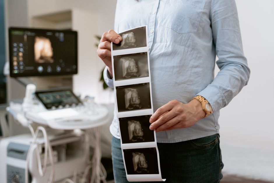
A PET scan image is a map of metabolic activity, not a photograph of your organs. The scanner detects a radioactive tracer that cells take up in proportion to how much energy they are using. Bright spots on the image mean high tracer uptake. Dark areas mean low uptake. Reading the image means comparing brightness across the body and against what is normal for each tissue.
That is the core skill. The rest is understanding why some tissues are always bright, why some bright spots are harmless, and why the number on the screen is not the same thing as a diagnosis.
What Does a PET Scan Image Actually Show?
A PET scan shows where cells are consuming the most glucose. The most common tracer is a radioactive form of glucose called fluorodeoxyglucose, or FDG. Cells pull it in through the same doorways they use for regular glucose. Once inside, the tracer gets trapped. The scanner then detects the radiation it emits.
Cancer cells often consume glucose faster than normal cells. That is the biological basis for using PET in oncology. But rapid glucose use is not unique to cancer. Infection, inflammation, healing tissue, and normal organs can all light up.
The image itself is usually fused with a CT scan. The CT provides anatomy — the shapes and locations of organs. The PET provides function — which of those structures are metabolically active. Reading a PET scan almost always means reading both together.
How Do You Read Uptake Values on a PET Scan?
The standardized uptake value, or SUV, is the number radiologists use to quantify brightness. It compares how much tracer concentrated in a spot versus how much would be there if the tracer were spread evenly through the whole body.
A higher SUV means more tracer uptake. But there is no universal cutoff that separates cancer from everything else. An SUV of 2.5 is often cited as a reference point in the medical literature, but it is not a diagnostic threshold. Some cancers have low uptake. Some benign conditions have high uptake.
Several factors shift the SUV up or down:
- Blood glucose level at the time of injection — high blood sugar competes with the tracer and can lower uptake
- Time between injection and scanning
- Body composition and weight
- The specific scanner and how it is calibrated
- Whether the patient moved during the scan
Because of this, SUVs are most useful when comparing the same patient’s scans over time, not when comparing one patient to another. A rising SUV in a known tumor is meaningful. An isolated SUV number in a new patient is a piece of a larger picture.
Why Are Some Normal Organs Always Bright on PET?
The brain, heart, kidneys, and bladder normally show high activity on an FDG PET scan. This is expected and does not indicate disease.
The brain uses glucose constantly and appears intensely bright. The heart muscle can take up a lot of tracer, though this varies — sometimes the heart is bright, sometimes it is not. The kidneys and bladder are bright because they are actively clearing the tracer from the body.
Other tissues show moderate, variable uptake. The liver usually has mild, even uptake. Bone marrow can be mildly active. Muscle uptake depends on recent activity — a patient who exercised or tensed up before the scan may show bright muscle.
Knowing what is normal is the foundation of reading any scan. A bright spot only means something when it appears where it should not.


What Causes False Positives and False Negatives?
False positives happen when something benign takes up a lot of tracer. Common causes include infection, inflammation, arthritis, healing wounds, and benign tumors. Brown fat — a type of fat that burns energy to produce heat — can also light up, especially in the neck and shoulders of thinner patients or in cold rooms.
False negatives happen when something serious does not take up much tracer. Some tumors are not very glucose-hungry. Small tumors may be below what the scanner can resolve. Certain cancer types, including some prostate cancers and some types of lung cancer, are known to be less reliably seen on FDG PET.
This is why PET is rarely read alone. A radiologist combines the PET findings with the CT images, the patient’s history, prior scans, and sometimes other tests. The scan informs a clinical judgment. It does not replace one.
How Do Radiologists Describe Metabolic Activity?
Radiologists use specific language to describe what they see. Understanding these terms helps you follow a report.
Hypermetabolic means increased metabolic activity — a bright spot. Hypometabolic means decreased activity. Diffuse means spread out over an area. Focal means concentrated in one spot. Physiologic means normal expected activity. Pathologic means activity that may indicate disease.
A report might say “focal hypermetabolic lesion in the right lung with SUV max of 6.2.” That means one concentrated bright spot in the right lung with a peak uptake value of 6.2. Whether that represents cancer depends on the full clinical context.
Reports often include a Deauville score when used for lymphoma. This is a five-point scale comparing tumor uptake to normal background tissues like the liver and mediastinum. It is a standardized way to describe response to treatment. It is specific to certain cancers and not used for all PET scans.
Can You Read a PET Scan Yourself?
You can understand the general principles, but you cannot reliably interpret your own scan. The image on the screen is a reconstruction from millions of detected events, processed by software. Reading it requires training in anatomy, pathology, and nuclear medicine.
What you can do is ask better questions. When you look at a PET report, useful questions include:
- What is the SUV max of the concerning area?
- Is the uptake focal or diffuse?
- Does it correspond to something visible on the CT portion?
- How does it compare to any previous scan?
- Could any benign condition explain this finding?
These questions help you have a more informed conversation with your doctor. They do not replace the doctor’s interpretation.
What Are the Limits of PET for Metabolic Activity?
PET is powerful but not unlimited. It shows glucose metabolism. It does not show everything happening in a cell. A tumor can be growing while showing low glucose uptake. An infection can mimic cancer. Inflammation after radiation therapy can create confusing results for weeks to months.
Some cancers are better imaged with other tracers. Prostate cancer, for example, is often evaluated with PSMA PET, which targets a protein on prostate cancer cells rather than glucose. Neuroendocrine tumors may use gallium-68 dotatate, which binds to somatostatin receptors. These are different tracers answering different questions.
Timing matters too. After surgery or radiation, inflammation can cause false positives. Most guidelines suggest waiting a certain period before scanning, but the ideal timing varies by situation and is not fully settled in every case.
How Does FDG Uptake Relate to Cancer Behavior?
Higher glucose uptake often correlates with more aggressive cancer, but the relationship is not absolute. Some aggressive cancers have modest uptake. Some slow-growing cancers can be quite bright.
What PET does well is show change over time. If a tumor’s uptake drops after treatment, that generally suggests the treatment is working. If uptake increases, that may suggest progression. This is why PET is often used to assess treatment response rather than to make an initial diagnosis alone.
The evidence for using PET to guide treatment decisions is strongest in certain cancers — lymphoma, lung cancer, and some others. In other cancers, the evidence is less clear. The role of PET continues to evolve as research accumulates.
Frequently Asked Questions
What does high uptake on a PET scan mean?
High uptake means cells in that area are consuming more glucose than surrounding tissue. It can indicate cancer, but infection, inflammation, and normal organ activity can also cause high uptake.
What is a normal SUV on a PET scan?
There is no single normal SUV value. Normal tissues have characteristic ranges, and what counts as concerning depends on the organ, the clinical context, and comparison with prior scans.
Can a PET scan tell the difference between cancer and inflammation?
Not always. Both cancer and inflammation can show increased glucose uptake. The CT portion of the scan, the shape of the finding, and the clinical history help distinguish them, but sometimes a biopsy is needed.
Why is the brain so bright on a PET scan?
The brain uses glucose continuously and at a high rate, so it always shows intense uptake on an FDG PET scan. This is normal and expected.
