
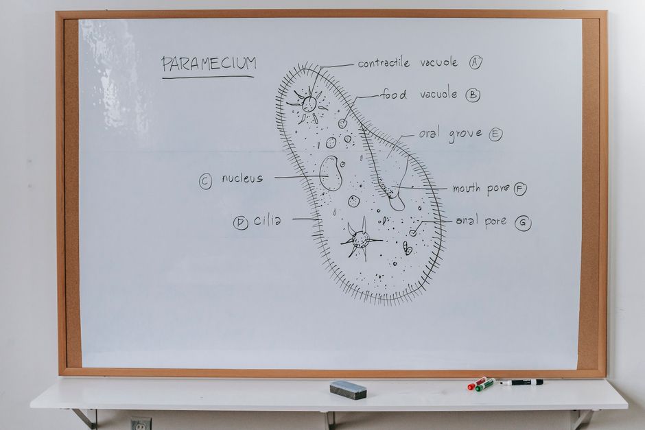
To draw a prokaryotic cell, start with an oval or rod shape for the cell envelope, then add a nucleoid (a region of DNA fibers) in the center, small dots for ribosomes, and thin lines for flagella or pili. Prokaryotic cells lack a nucleus and membrane-bound organelles, so your drawing should show the genetic material floating freely in the cytoplasm. Follow these steps to create an accurate biological diagram of a bacterium or archaeon.
What You Need to Know Before Drawing a Prokaryotic Cell
Prokaryotes are single-celled organisms without a nucleus. Bacteria and archaea are the two main groups. Their cells are generally smaller than eukaryotic cells and have a simpler internal structure. The key parts to include in a drawing are the cell wall, cell membrane, cytoplasm, nucleoid (DNA), ribosomes, and sometimes flagella, pili, and a capsule. A good diagram is both accurate and neatly labeled.
Most textbooks show a typical bacterial cell as a short rod (bacillus) or a sphere (coccus). For a step-by-step guide, a rod shape is easier to draw and clearly shows internal components.
Step 1: Draw the Outer Layers (Capsule, Cell Wall, Cell Membrane)
Begin with an oval or slightly elongated shape. This represents the entire cell envelope. Draw a thin outer line to represent the capsule – a protective layer of polysaccharides. Not all prokaryotes have a capsule, but including it shows a common structure.
Inside the capsule, add a thicker line for the cell wall. The cell wall gives the cell its shape and prevents it from bursting. For most bacteria, this wall contains peptidoglycan. Inside the cell wall, draw a dashed or double line for the cell membrane. The membrane is a phospholipid bilayer that controls what enters and leaves the cell.
Together, these three layers form the protective envelope. Leave a clear space between them in your drawing so each layer is distinguishable.
Step 2: Add the Internal Structures (Nucleoid, Ribosomes, Plasmids)
Inside the cell, draw a large, irregular, wavy line or a cluster of squiggly lines in the center. This is the nucleoid – the region where the single circular chromosome is located. Unlike a eukaryotic nucleus, the nucleoid has no surrounding membrane.
Scatter small dots throughout the cytoplasm. These are ribosomes. Prokaryotic ribosomes are smaller (70S) than eukaryotic ones, but in a diagram they look like tiny circles or dots. They are not membrane-bound.
Optional but helpful: draw one or two small circles separate from the nucleoid to represent plasmids. Plasmids are small, circular pieces of extra DNA that can carry genes for antibiotic resistance or other traits. They float freely in the cytoplasm.




Step 3: Draw Appendages (Flagella, Pili, Fimbriae)
Flagella are long, whip-like structures that help the cell move. Draw one or two thin, wavy lines coming from one end of the cell. Each flagellum should have a curved shape, not a straight line. In some bacteria, flagella cover the entire surface; in others, they are at one pole.
Pili are shorter, straighter, hair-like projections. They help cells attach to surfaces or other cells. Draw several short, straight lines around the cell’s outer surface. Fimbriae are even shorter and more numerous – you can show them as a fuzzy coat around the outside of the cell wall, but only if your diagram requires that level of detail.
Step 4: Label Your Drawing
Use clear, straight lines with arrows pointing to each structure. Label these parts: capsule (if included), cell wall, cell membrane, cytoplasm, nucleoid, ribosomes, flagellum (or flagella), and pilus (or pili). If you added plasmids, label those too. Use a ruler for the label lines to keep the diagram neat.
Write each label in print letters, not cursive, so it is easy to read. Put the labels outside the drawing and keep lines parallel when possible. This makes your diagram look professional and helps teachers or readers identify each part quickly.
Common Mistakes When Drawing Prokaryotic Cells
One frequent error is drawing a nucleus. Prokaryotes do not have a nucleus. Instead of a round, membrane-bound nucleus, show the DNA as a tangled region (nucleoid). Another mistake is making the cell wall look like a single line – it should be drawn as a distinct layer between the capsule and membrane.
Some students forget to include ribosomes or label them. Ribosomes are essential for protein synthesis and are present in all prokaryotic cells. Also, do not draw mitochondria, chloroplasts, or other membrane-bound organelles – those belong in eukaryotic cells only.
Finally, check that the flagella are attached to the cell wall or membrane, not floating separately. They are anchored at the base by a protein structure called the basal body.
Frequently Asked Questions
Should I draw a nucleus in a prokaryotic cell?
No. Prokaryotic cells do not have a nucleus. Draw the DNA as a nucleoid – an irregular region without a membrane.
How many flagella should I draw?
It depends on the species. Most drawing assignments use one or two flagella to show the structure. Check your specific instructions.
Do I need to include every labeled part listed in my textbook?
Include only the parts that your assignment or teacher requires. Common essentials are cell wall, cell membrane, nucleoid, ribosomes, and flagella.
Can I use color in my prokaryotic cell diagram?
Yes. Many textbooks use color – for example, blue for the nucleoid, green for the cell wall, red for membrane. Use consistent colors and add a color key.
