
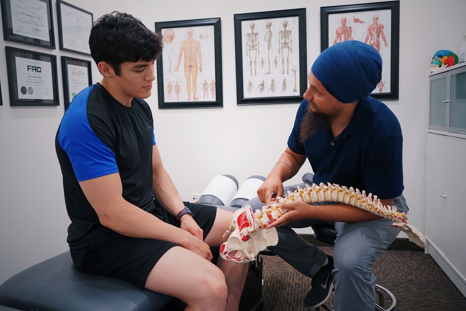
Diagnosing a spinal cord injury requires a combination of emergency imaging, a detailed neurological exam, and specific tests to measure nerve function. Doctors typically start with a CT scan or MRI in the emergency room to see the bones and soft tissues of the spine. They also perform a physical exam to check your ability to move, feel sensation, and control reflexes. The goal is to find the exact location and severity of the injury as fast as possible because early treatment can prevent permanent damage.
What Happens First When a Spinal Cord Injury Is Suspected?
The first step happens before you even reach the hospital. Emergency medical technicians (EMTs) stabilize your head and neck to prevent any movement. They use a rigid collar and a backboard. This is critical because moving an unstable spine can turn a partial injury into a complete one.
Once you arrive at the trauma center, the emergency team takes over. They follow a standard protocol called Advanced Trauma Life Support (ATLS). This means they check your airway, breathing, and circulation first. Only after you are stable do they focus on the spine. The team will ask about the mechanism of injury — a car crash, a fall, or a sports accident. They also need to know if you lost consciousness or felt any numbness or tingling at the scene.
Research published in the Journal of Neurosurgery: Spine shows that the time from injury to spinal stabilization is one of the strongest predictors of recovery. Every hour matters.
Which Imaging Tests Are Used to Diagnose a Spinal Cord Injury?
Imaging is the backbone of diagnosis. The first test is almost always a CT scan. A CT scan is fast — it takes about five minutes. It gives doctors a clear picture of the bones in your spine. It can show fractures, dislocations, or bone fragments pressing on the spinal cord. The American College of Radiology recommends CT as the first-line imaging for trauma patients with suspected spinal injury.
If the CT is unclear or if doctors suspect damage to the spinal cord itself, they order an MRI. An MRI shows soft tissues, including the spinal cord, nerves, discs, and ligaments. It can reveal swelling, bleeding, or a herniated disc that is compressing the cord. An MRI takes longer — usually 30 to 45 minutes — and requires you to lie completely still. The table below compares the two main imaging methods.
| Test | What It Shows | Time to Complete | Best For |
|---|---|---|---|
| CT Scan | Bone fractures, dislocations, alignment | 5 minutes | Emergency trauma evaluation |
| MRI | Spinal cord, nerves, discs, ligaments, bleeding | 30–45 minutes | Assessing cord compression and soft tissue damage |
X-rays are sometimes used but are less reliable. A plain X-ray can show a fracture or misalignment, but it misses many injuries that a CT would catch. Most trauma centers skip X-rays and go straight to CT.
How Do Doctors Test Nerve Function and Reflexes?
After imaging, the next step is a thorough neurological exam. This is not a simple check. It is a systematic evaluation of motor strength, sensation, and reflexes at every level of the spine. Doctors use a standardized scale called the International Standards for Neurological Classification of Spinal Cord Injury (ISNCSCI).
The exam tests 10 key muscle groups on each side of your body. For example, bending your elbow tests the C5 nerve root. Lifting your thigh tests L2. Each muscle is graded from 0 (no movement) to 5 (normal strength). Sensation is tested with a light touch and a pinprick at 28 specific spots on each side of the body. This creates a sensory map that pinpoints the level of injury.
Reflexes are also checked. A brisk reflex below the injury level can indicate an upper motor neuron injury. Absent reflexes suggest damage to the lower motor neurons or nerve roots. The combination of these findings tells doctors whether the injury is complete or incomplete. An incomplete injury means some nerve signals still get through below the injury level. A complete injury means no signals pass at all.




What Are Electrodiagnostic Tests and When Are They Used?
Electrodiagnostic tests measure the electrical activity of nerves and muscles. They are not used in the emergency room. They are done days or weeks later when the patient is stable. These tests help confirm the diagnosis and predict recovery.
The two main tests are electromyography (EMG) and nerve conduction studies (NCS). EMG uses a thin needle electrode inserted into muscles to record electrical activity at rest and during contraction. NCS uses small electrical shocks on the skin to measure how fast nerves send signals. If a nerve is damaged, the signal travels more slowly or not at all.
Research from the American Association of Neuromuscular and Electrodiagnostic Medicine indicates that EMG and NCS can detect nerve root avulsion — where a nerve is torn from the spinal cord. This is a severe injury that does not heal on its own. Finding it early changes the treatment plan. Some patients may need nerve transfer surgery.
These tests are not painful for most people, though you may feel a mild pinch from the needle or a brief shock from the NCS. Results come back within a few days.
How To Diagnose a Spinal Cord Injury Tests Exams in Special Situations
Some patients cannot participate in a standard neurological exam. A person who is unconscious, sedated, or has a traumatic brain injury cannot tell you where they feel a pinprick. In these cases, doctors rely on other methods.
One option is somatosensory evoked potentials (SSEPs). This test stimulates a nerve in the wrist or ankle and measures the electrical signal as it travels up the spinal cord to the brain. It does not require the patient to respond. Another option is motor evoked potentials (MEPs), which stimulate the brain and measure the response in the muscles. These tests can be done at the bedside in the intensive care unit.
A systematic review in Critical Care Medicine found that SSEPs and MEPs can predict neurological recovery in unconscious patients with spinal cord injuries with about 85 percent accuracy. This is not perfect, but it gives families and doctors useful information during the first days when the patient cannot be examined normally.
For children, the approach is similar but adjusted. Children’s spines are more flexible, so they can have a spinal cord injury without a fracture. This is called SCIWORA — spinal cord injury without radiographic abnormality. It is more common in kids under eight. If a child has neurological symptoms but a normal CT scan, doctors will still order an MRI to look for ligament damage or cord swelling.
What Are Common Misconceptions About Spinal Cord Injury Diagnosis?
A common myth is that a normal CT or X-ray means the spinal cord is fine. This is false. As mentioned above, SCIWORA exists. Also, conditions like spinal cord contusion or central cord syndrome can show up on MRI but not on CT. If you have symptoms like numbness, weakness, or loss of bladder control, you need an MRI even if the CT looks clean.
Another misconception is that a complete injury means you will never recover any function. This is not entirely accurate. The term “complete” means no nerve signals pass the injury site at the time of testing. But some patients with a complete injury regain some function over months, especially if the cord was not severed. Recovery depends on the type and severity of damage, not just the label.
Some people also believe that you can “walk off” a spinal injury if you can move your legs. This is dangerous. A spinal cord injury can cause swelling that gets worse over hours. You might be able to move right after the accident but lose function later. That is why paramedics immobilize everyone with a suspected spine injury until imaging rules it out.
Frequently Asked Questions
How long does it take to diagnose a spinal cord injury?
A CT scan can be done within minutes of arrival at the hospital, and a preliminary diagnosis is often made in the first hour. A full diagnosis with MRI and neurological exam may take several hours.
Can a spinal cord injury be missed on a CT scan?
Yes, a CT scan can miss injuries to the spinal cord itself, such as swelling or bruising. An MRI is needed to see soft tissue damage that a CT cannot detect.
What is the ASIA scale for spinal cord injury?
The ASIA scale is a standardized system that grades injury severity from A (complete injury) to E (normal function). It is based on motor and sensory testing at specific points on the body.
Do you need to be awake for a spinal cord injury exam?
No, doctors can use evoked potential tests to assess nerve function in patients who are unconscious or sedated. These tests do not require the patient to respond.
