
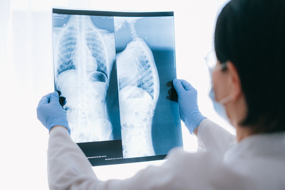
A lung infection is diagnosed by combining the story of your illness with a physical exam and, usually, at least one test. The most common first test is a chest X-ray, which can show areas of inflammation in the lungs. When more detail is needed, clinicians add sputum cultures, blood tests, or a CT scan. The right combination depends on how sick you are, your history, and what the exam suggests.
How Do Doctors Tell If You Have a Lung Infection?
Diagnosis starts with a conversation, not a machine. A clinician asks how long you have been sick, whether your cough is dry or brings up mucus, and whether you have fever, chills, or trouble breathing. That history narrows the field quickly.
Next comes a physical exam. The clinician listens to your lungs with a stethoscope, checking for crackles, wheezing, or sounds that are muffled in one area. They may tap on your chest and check your oxygen level with a small device on your finger called a pulse oximeter. They also check your breathing rate and heart rate.
No single finding confirms a lung infection on its own. A normal-sounding chest does not rule one out, especially in older adults. Fever can be absent in people over 65 even when pneumonia is present. This is why the history and exam guide which tests to order rather than standing alone as proof.
What Is the Difference Between Pneumonia, Bronchitis, and Other Lung Infections?
Lung infections are grouped by where they sit in the airways. That location shapes both symptoms and testing.
Bronchitis affects the large airways, called the bronchi. It usually causes a cough, often with mucus, and may follow a cold. Chest X-rays typically look normal in acute bronchitis, which is one reason imaging is not always ordered for a mild cough.
Pneumonia affects the air sacs deep in the lungs. Those sacs can fill with fluid and inflammatory cells, which is what shows up as a shadow or opacity on a chest X-ray. Pneumonia tends to bring fever, chills, cough, and shortness of breath.
Other infections include bronchiolitis, which affects the smallest airways and is most common in infants, and lung abscess, a pocket of pus in the lung tissue. Tuberculosis is a bacterial infection with its own distinct testing path. Fungal lung infections are less common in healthy people but occur more often in those with weakened immune systems or in specific geographic regions.
The distinction matters because treatment differs. Bacterial pneumonia may need antibiotics. Viral infections do not respond to antibiotics. Knowing which one you have changes what happens next.
What Tests Are Used To Diagnose a Lung Infection?
Testing ranges from a simple X-ray to procedures that sample lung tissue. Most people need only one or two.
Chest X-Ray
The chest X-ray is the standard first imaging test for suspected pneumonia. It is fast, widely available, and low in radiation. It can reveal fluid buildup or areas of inflammation. A limitation: early in an infection, an X-ray can look normal. A repeat image a day or two later sometimes shows changes that were not visible at first.
Pulse Oximetry and Blood Gas Tests
A pulse oximeter clips onto your finger and estimates how much oxygen your blood is carrying. A normal reading is generally 95% or higher at sea level, though this varies with altitude and other factors. If the reading is low or the clinician needs exact numbers, they may draw blood from an artery to measure oxygen and carbon dioxide directly. This is called an arterial blood gas test.
Sputum Culture
If you can cough up mucus, a sample can be sent to a lab. There, technicians try to grow the organism causing the infection and test which antibiotics work against it. This takes time — often a day or more — so treatment usually starts before results come back. Not everyone can produce a good sample, and the results are not always conclusive.
Blood Tests
A complete blood count can show whether white blood cells are elevated, which sometimes supports a bacterial cause, though it does not confirm one. Blood cultures can detect bacteria that have entered the bloodstream. A test called procalcitonin is sometimes used to help judge whether a bacterial infection is likely, though its role varies by setting and it is not a stand-alone answer.
CT Scan
A CT scan gives a much more detailed picture than a chest X-ray. It is usually reserved for cases where the X-ray is unclear, the infection is not improving, or the clinician suspects a complication such as an abscess or a blockage. CT uses more radiation than a plain X-ray, so it is not a routine first step.
Bronchoscopy
In a bronchoscopy, a thin flexible tube is passed through the mouth or nose into the airways. It allows the clinician to see inside and collect samples directly. This is used when simpler tests have not given an answer, when a person is very ill, or when the immune system is weakened.
Other Tests
When tuberculosis is suspected, a skin test or a blood test is used, along with sputum analysis. For suspected fungal infections, specific blood or sputum tests may be ordered. Rapid tests for viruses such as influenza or respiratory syncytial virus (RSV) can identify a viral cause, which changes treatment.





How Do Doctors Choose Which Tests To Order?
Testing is not one-size-fits-all. Clinicians weigh several factors before deciding what to order.
- How sick you are. A mild cough in an otherwise healthy person may need no imaging at all. Trouble breathing, low oxygen, or confusion pushes testing forward quickly.
- Your age and health history. Older adults and people with chronic lung disease, heart disease, or diabetes may need closer evaluation.
- Your immune status. People with weakened immune systems can get infections from organisms that rarely affect others, so broader testing is often used.
- Where you were exposed. Travel or residence in certain regions raises the chance of specific fungal or tuberculosis infections.
- Whether you are improving. If symptoms are not getting better as expected, that is a reason to look further.
A tool some clinicians use is a severity score, such as the CURB-65, which factors in confusion, blood urea nitrogen, breathing rate, blood pressure, and age. It helps estimate whether a person can be treated at home or needs hospital care. It is a guide, not a verdict.
Why Diagnosis Is Not Always Straightforward
Lung infections can look like other conditions. Asthma, heart failure, and even some cancers can cause cough, shortness of breath, and chest X-ray changes that resemble pneumonia. This overlap is a real reason testing sometimes expands.
Test results can also be misleading. A sputum sample may grow bacteria that are just colonizing the airway rather than causing disease. An X-ray read as clear early on may show an infection a day later. And some people, particularly those over 65, may have a lung infection without fever or a productive cough.
Because of this, diagnosis is a process of assembling clues rather than reading a single result. The clinician weighs the whole picture — symptoms, exam, imaging, and lab work — before settling on a cause and a plan.
When Should You Seek Medical Care for Suspected Lung Infection?
Some symptoms call for prompt evaluation. Seek care if you have trouble breathing, chest pain, a fever that will not go down, confusion, or lips or fingertips that look bluish. A cough that lasts more than a few weeks, or one that brings up blood, also warrants a medical visit.
For most people with a mild cough and no warning signs, watching for a few days is reasonable. But the threshold for seeking care is lower in infants, older adults, pregnant people, and anyone with a chronic illness or weakened immune system. If you are unsure, a call to a clinician is a reasonable step.
No test can be interpreted in isolation. The value of any single result depends on the context of your symptoms and exam. That is why the diagnosis is made by a person, not a machine.
Frequently Asked Questions
What is the most common test for a lung infection?
A chest X-ray is the most common first imaging test when pneumonia is suspected. It can show areas of inflammation or fluid in the lungs, though it may look normal very early in an infection.
Can a blood test diagnose pneumonia?
Blood tests can support the diagnosis but do not confirm it on their own. A complete blood count may show elevated white blood cells, and blood cultures can detect bacteria in the bloodstream, but the full picture comes from symptoms, exam, and imaging together.
How long does it take to get lung infection test results?
A chest X-ray result is usually available within hours. Sputum and blood cultures take longer, often a day or more, because the lab must grow the organism before it can be identified.
Do you always need a chest X-ray for a lung infection?
No. A mild cough in an otherwise healthy person may be managed without imaging. A chest X-ray is more likely when symptoms are severe, not improving, or when the exam suggests pneumonia.
