
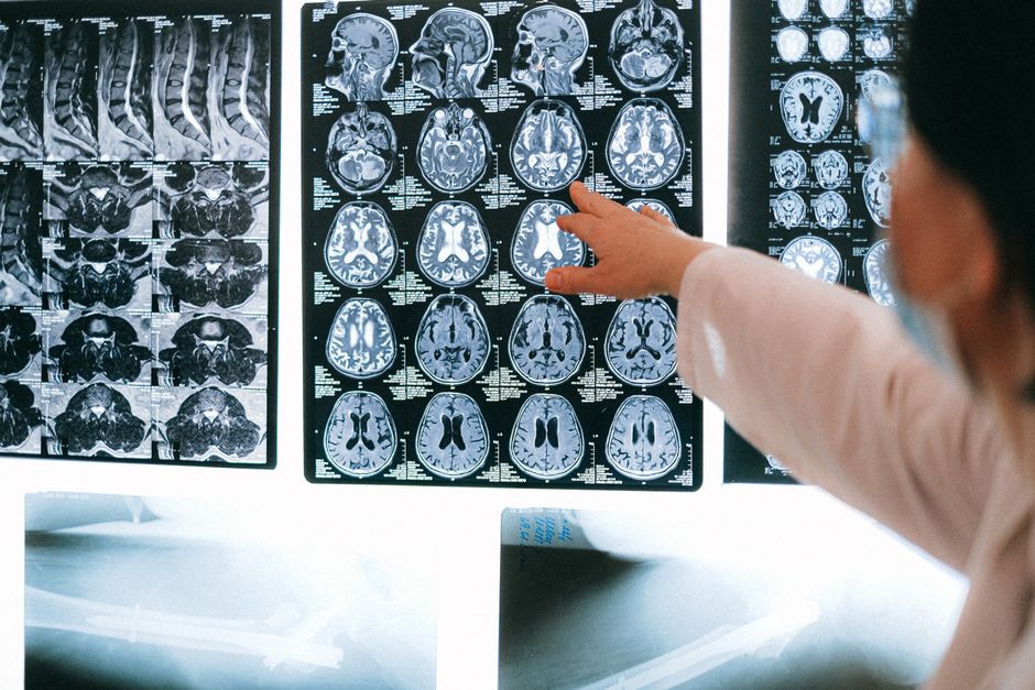
When you have a suspicious lump, an abnormal blood test, or an unclear finding on a scan, two very different medical specialties often end up working the same case. Radiology uses imaging — X-rays, CT scans, MRIs, ultrasounds — to see inside the body without cutting it open. Pathology examines tissue, blood, and other samples under a microscope and through lab tests to identify what is actually happening at the cellular level. Together, they answer two connected questions: where is the problem, and what is it?
Neither specialty is a backup for the other. They answer different questions, and the diagnosis usually gets firmer when both answers line up.
What Does Radiology Do That Pathology Cannot?
Radiology shows structure and function in a living person, in real time, without removing anything. That is something pathology simply cannot do.
A radiologist reads images to locate a problem, measure its size, and describe its shape and borders. They can see whether a mass is solid or fluid-filled, whether it has spread to nearby lymph nodes, and how it sits relative to organs and blood vessels. Imaging also tracks change over time — a nodule that stays the same size for two years tells a different story than one that has doubled.
What radiology generally cannot do is tell you with certainty what cells make up a mass. A CT scan can show a spot on a lung with great precision. It cannot confirm whether those cells are cancerous, inflamed, or scar tissue. That distinction usually requires tissue.
There are exceptions worth knowing. Some imaging findings are so characteristic that radiologists can diagnose with high confidence without a biopsy — certain benign bone growths, simple fluid-filled cysts, or classic patterns of a healed fracture. But for most suspected cancers, imaging alone is not considered definitive.
What Does Pathology Add That Imaging Misses?
Pathology provides the tissue-level answer: what the cells are, whether they are benign or malignant, and — if malignant — what type of cancer it is and how aggressive it appears.
A pathologist examines tissue samples under a microscope and often runs additional tests on the same sample. These can include special stains that highlight specific proteins, and molecular tests that look for gene changes in the tumor. That information can shape which treatments are likely to work. Two tumors that look identical on a scan can behave very differently based on their molecular profile — and only pathology can reveal that difference.
Pathology also goes beyond cancer. It identifies infections in tissue, confirms inflammatory diseases, and checks whether a transplanted organ is being rejected. Blood and body fluid tests run through pathology labs diagnose everything from anemia to diabetes to infections.
The trade-off is that pathology usually requires a sample. Getting that sample means a biopsy, a procedure, or surgery — each with its own risks, waiting time, and cost.
How Radiology And Pathology Work Together In Diagnosis
The two specialties work as a relay team, and the handoff matters as much as either runner.
The process usually follows a sequence:
- Imaging first. A scan identifies an abnormality and describes where it is and what it looks like.
- Biopsy planning. The radiologist uses the images to guide a needle to the exact spot — this is called image-guided biopsy. Ultrasound, CT, and sometimes MRI or mammography are used to steer the needle precisely.
- Tissue analysis. The pathologist examines the sample and issues a diagnosis, often with details about tumor type and grade.
- Correlation. The radiologist and pathologist — often with the treating physician — compare findings. Does the tissue diagnosis fit the imaging appearance? When they disagree, that mismatch is a signal to look again.
That last step is where the real value sits. A biopsy result that does not match the imaging picture can mean the needle missed the target, the sample came from the wrong area, or the imaging interpretation needs revisiting. Radiologists and pathologists flag these discrepancies, and the case often gets reviewed again before treatment decisions are made.
Image-guided biopsy is a good example of the partnership in action. Without imaging, a surgeon might need open surgery just to find the target. With imaging guidance, a needle sample can often be taken through the skin with far less disruption. The pathologist then gets a small but precisely located piece of tissue — and the precision comes entirely from the radiologist’s images.
Why Do Doctors Sometimes Need Both a Scan and a Biopsy?
Because each answers a question the other cannot, and treatment decisions often need both answers.
Imaging shows the full extent of disease — whether a tumor has spread, how large it is, and what organs are involved. That is called staging, and it shapes the overall treatment plan. Pathology shows what the tumor is made of and how it is likely to behave. That is called characterization, and it shapes which specific treatments make sense.
A patient might have a lung mass that looks malignant on CT. The scan tells the team it is confined to one area and has not spread. The biopsy tells them it is a type of cancer that responds to particular targeted drugs. Neither piece of information alone would be enough to plan treatment well.
There are situations where a biopsy is not needed. If imaging is highly characteristic and the treatment would be the same regardless, some clinicians proceed without tissue confirmation. This happens more often in certain benign conditions or in patients for whom biopsy carries high risk. It is a judgment call, not a rule, and it varies by condition and by clinician.
What Happens When Radiology and Pathology Disagree?
A mismatch between imaging and tissue findings is not rare, and it usually triggers a closer look rather than a final answer.
Disagreement can happen for several reasons. The biopsy needle may have sampled tissue next to the actual lesion rather than the lesion itself. The imaging finding may have been over- or under-interpreted. Or the condition may genuinely look different under the microscope than it does on a scan — some inflammatory conditions mimic tumors on imaging, and some tumors look deceptively benign.
When this happens, the case often goes to a multidisciplinary review, where radiologists, pathologists, and treating physicians look at the images and slides together. Additional sampling, repeat imaging, or specialized testing may follow. This kind of review is standard practice in cancer centers and is one reason tumor boards exist.
The mismatch itself is useful information. It tells the clinical team that something in the picture is incomplete, and it prevents premature treatment decisions based on partial data.
How Has Technology Changed the Partnership?
Digital imaging and digital pathology have made it easier for both specialties to look at the same case at the same time.
Pathology slides can now be scanned into high-resolution digital images, which means a pathologist can review a case remotely and share it instantly with colleagues. Radiology images have been digital for years. Together, this allows faster consultation and second opinions without shipping physical slides or films.
Artificial intelligence tools are being developed for both fields — to help flag suspicious areas on scans and to assist pathologists in spotting abnormal cells. The evidence for these tools is still developing. Some studies suggest they can improve detection in specific settings, but they are not a replacement for trained specialists, and their real-world impact on patient outcomes is still being studied.
What has not changed is the core logic. Imaging locates and characterizes the problem in the body. Pathology identifies what it actually is at the cellular level. The diagnosis gets stronger when both agree — and more careful when they do not.
Frequently Asked Questions
Can a radiologist diagnose cancer without a biopsy?
In some cases, imaging findings are characteristic enough that radiologists can diagnose with high confidence, but for most suspected cancers a tissue sample is still needed for a definitive diagnosis. Imaging alone generally cannot confirm what cells make up a mass.
What is an image-guided biopsy?
It is a procedure where a radiologist uses ultrasound, CT, or another imaging method to guide a needle to the exact location of an abnormality. This allows a tissue sample to be taken precisely, often without open surgery.
Why would a doctor order both a CT scan and a biopsy?
A CT scan shows where a problem is and how far it extends, while a biopsy shows what the tissue actually is. Treatment planning often needs both pieces of information.
What does it mean if the imaging and biopsy results don’t match?
It usually means the case needs closer review, often by a team of radiologists, pathologists, and treating physicians. A mismatch can happen if the biopsy missed the target or if the condition looks different on imaging than under a microscope.
