
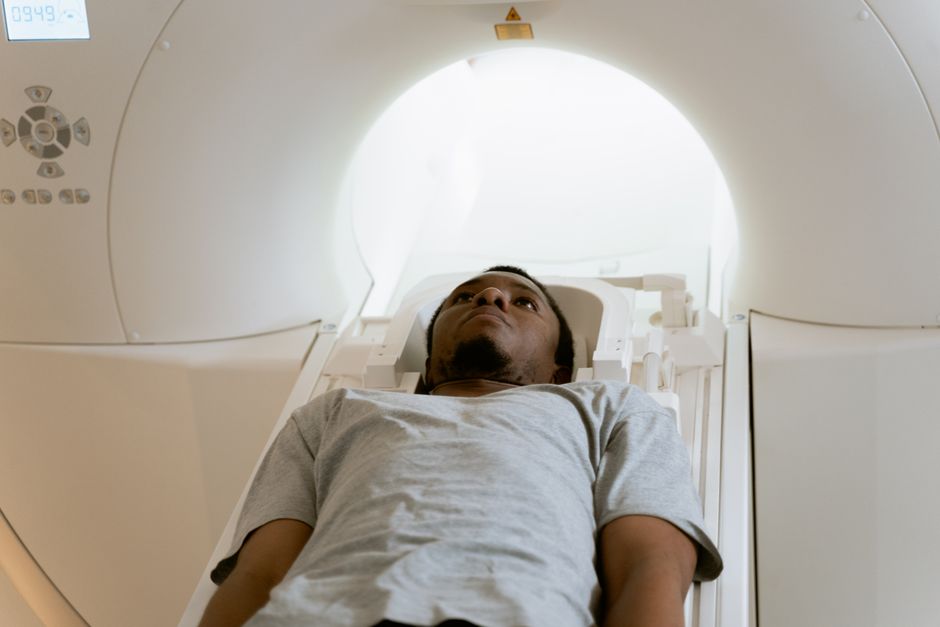
A CT scan delivers a radiation dose roughly equivalent to what you would naturally absorb from the environment over one to five years. The exact amount depends on the body part being scanned, the machine used, and your size. For most single scans, the dose ranges from 1 to 10 millisieverts (mSv), though the most common average is around 7 mSv for an abdominal scan.
What Does the Radiation Number Actually Mean?
Radiation dose is measured in millisieverts (mSv). This unit accounts for both the amount of energy absorbed and how different tissues respond to radiation. It is the standard way doctors and physicists compare imaging exams.
A single chest CT delivers about 7 mSv. A head CT is lower, around 2 mSv. A coronary artery calcium scan for heart health is roughly 1.5 mSv. These figures come from standard reference data used by medical physicists and radiology organizations.
To put this in perspective, the average American receives about 3 mSv per year from natural background radiation. This comes from radon in soil, cosmic rays at high altitudes, and trace radioactive elements in food and water. A single abdominal CT therefore exposes you to roughly two to three years of natural background radiation in a few minutes.
How Does a CT Scan Compare to Other Imaging Tests?
Not all imaging uses radiation. Ultrasound and MRI use sound waves and magnetic fields respectively, delivering no ionizing radiation at all. X-rays and CT scans both use ionizing radiation, but the doses differ significantly.
- Dental X-ray: about 0.005 mSv
- Chest X-ray: about 0.1 mSv
- Mammogram: about 0.4 mSv
- Head CT: about 2 mSv
- Chest CT: about 7 mSv
- Abdominal CT: about 8 mSv
A CT scan is essentially a series of X-ray images taken from multiple angles and combined by computer into cross-sectional slices. Because it gathers far more information than a single X-ray, it delivers a much higher dose.
Why Do Repeated CT Scans Raise Concern?
Radiation damage to cells accumulates over a lifetime. Each exposure adds a small chance of future cancer. The risk is not zero, but for a single scan it remains very small.
Research consistently shows that the risk becomes more meaningful with cumulative exposure. Studies of atomic bomb survivors and radiation workers provide the foundation for understanding this relationship. These populations received higher doses than typical medical imaging, so scientists extrapolate downward to estimate risk at CT levels.
The estimated additional lifetime cancer risk from one 10 mSv scan is roughly 1 in 2,000 for an adult. That is a 0.05 percent increase over the baseline cancer risk every person already carries. For context, the lifetime risk of developing any cancer is about 39 percent for men and 38 percent for women in the United States.
Children are more sensitive to radiation than adults. Their cells divide more rapidly, and they have more years of life ahead for damage to manifest. This is why pediatric imaging protocols use lower doses and why doctors are especially cautious about scanning children.





When Is a CT Scan Worth the Radiation?
A CT scan is one of the most powerful diagnostic tools in medicine. It can detect internal bleeding after trauma, identify blood clots in the lungs, evaluate appendicitis, and guide cancer staging. In many emergency situations, the benefit of an accurate diagnosis far outweighs the small radiation risk.
Doctors follow the principle of ALARA — As Low As Reasonably Achievable. This means they order CT scans only when the clinical question cannot be answered by safer methods. If an ultrasound or MRI can provide the same answer, most physicians will choose those first.
You should not refuse a medically necessary CT scan out of radiation fear. The risk of missing a serious condition like a pulmonary embolism or a brain bleed is typically far greater than the cancer risk from the scan itself. The conversation about radiation is about avoiding unnecessary scans, not avoiding necessary ones.
How Do Hospitals Keep CT Doses Low?
Modern CT machines have built-in dose reduction technology. Automatic exposure control adjusts the radiation output based on your body size and the density of the tissue being imaged. A smaller patient automatically receives a lower dose than a larger patient.
Newer scanning protocols use iterative reconstruction, a computer algorithm that produces clearer images with less radiation. Some newer systems also use tin filtration, which removes low-energy X-rays that contribute to dose without improving image quality.
Hospitals track radiation doses for every patient. Accredited imaging centers must meet dose benchmarks set by professional organizations. If you have had many CT scans in the past, you can ask your doctor whether a facility that participates in the American College of Radiology Dose Index Registry performs your scan. This registry tracks whether facilities stay within expected dose ranges.
You can also ask a simple question before any scan: “Has this test been discussed with a radiologist?” Radiologists are physicians specially trained in imaging appropriateness. Many hospitals require radiology input before CT scans are performed, but not all do.
What About Contrast Dye and Radiation?
Many CT scans use intravenous contrast dye to make blood vessels and organs more visible. This dye contains iodine and does not add radiation dose. The radiation comes only from the X-ray beam itself.
Contrast dye carries its own separate risks, including allergic reactions and kidney strain in people with pre-existing kidney disease. These risks are uncommon but real. If you have diabetes, high blood pressure, or known kidney problems, tell your doctor before receiving contrast.
The contrast discussion is separate from the radiation discussion. Both matter, but they are different considerations. A scan with contrast does not expose you to more radiation than the same scan without it.
How Can You Track Your Cumulative Radiation Exposure?
You have no national registry tracking your medical radiation. Your imaging records exist only in the facilities where your scans were performed. This makes it easy to lose track of how many scans you have received over a lifetime.
Keep a personal record. Write down the date, type of scan, body part, and facility for every imaging exam you receive. If the report lists the dose in mSv, record that number too. Many modern reports include this information, though not all do.
If you have had multiple CT scans, you can ask your primary care doctor to review the cumulative record. Some patients who have received many scans over years may benefit from discussing whether future imaging could use alternative methods. This is not a standard recommendation for everyone — it applies mainly to people with chronic conditions requiring frequent imaging.
Frequently Asked Questions
How much radiation is in one CT scan?
A typical CT scan delivers between 1 and 10 mSv depending on the body part, with abdominal scans averaging around 8 mSv and head scans around 2 mSv.
How many CT scans are safe in a year?
No official limit exists because medical necessity determines whether each scan is appropriate, not a calendar count.
Does CT scan radiation stay in your body?
No, the radiation passes through your body during the scan and does not remain afterward.
Is a CT scan more radiation than an X-ray?
Yes, a CT scan delivers significantly more radiation than a single X-ray, typically 50 to 100 times more depending on the specific exams compared.
