
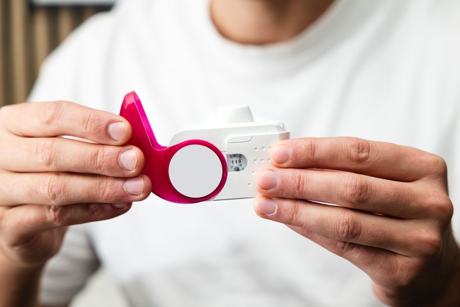
Most people breathe without thinking about it. COPD changes that. The diagnosis usually starts with a simple breathing test called spirometry, which measures how much air you can blow out and how fast. Imaging such as a chest X-ray or CT scan can support the diagnosis, rule out other problems, and show changes in lung structure. Together, these tools help a clinician confirm COPD and understand how severe it is.
How Is COPD Diagnosed With Spirometry, Imaging, and More?
Spirometry is the test that confirms COPD. A diagnosis requires evidence of airflow obstruction that does not fully reverse after a bronchodilator medicine.
During spirometry, you take a deep breath and blow out as hard and as long as you can into a tube connected to a machine. The test reports two key numbers. FEV1 is the volume of air you can force out in one second. FVC is the total volume you can force out in one full breath.
The ratio of FEV1 to FVC is the number that matters most for diagnosis. In COPD, this ratio falls below 0.70 after bronchodilator use. That threshold is well established in clinical guidelines and is used worldwide to define persistent airflow obstruction.
Spirometry is not a perfect test. It requires effort and coordination, and results can vary if the test is not done properly. Clinicians usually ask for at least three acceptable blows to make sure the numbers are reliable. Poor technique can produce misleading results in either direction.
Imaging plays a supporting role, not a diagnostic one. A chest X-ray cannot confirm COPD on its own. It can show hyperinflation, flattened diaphragms, or a enlarged heart shadow, and it helps exclude other causes of breathlessness such as heart failure or pneumonia. A CT scan shows structural damage in more detail, including emphysema and airway thickening, and can help identify other conditions.
What Symptoms and History Point Toward COPD?
COPD should be considered in anyone with ongoing breathlessness, a long-standing cough, or sputum production, especially with a history of smoking or other inhaled exposures.
The classic symptoms are breathlessness that worsens over time, a cough that does not go away, and phlegm. Many people first notice breathlessness during exertion — climbing stairs, walking uphill, or carrying groceries. They often attribute it to aging or being out of shape. That delay is common and can mean the disease is more advanced by the time it is diagnosed.
Wheezing and chest tightness can also occur. Some people have frequent chest infections. A clinician will ask about smoking history, occupational exposures such as dust or fumes, and family history. They will also ask about other conditions that can cause similar symptoms.
One point that is often missed: not everyone with COPD has smoked. A significant minority develop the disease from other exposures or genetic factors. Never assume the diagnosis is ruled out just because someone never smoked.
What Other Tests Help Confirm or Rule Out COPD?
Several tests support the diagnosis, measure severity, and rule out conditions that mimic COPD.
- Post-bronchodilator spirometry — the confirmatory test. It distinguishes COPD from asthma, where airflow obstruction often reverses substantially after a bronchodilator.
- Chest X-ray — helps exclude other causes of symptoms and may show signs of hyperinflation.
- CT scan — shows emphysema and airway changes in detail; used when the diagnosis is uncertain or when surgery or other procedures are being considered.
- Pulse oximetry and arterial blood gas — measure oxygen levels; blood gas testing is used in more advanced disease or during flare-ups.
- Alpha-1 antitrypsin testing — recommended for certain patients, particularly those with early-onset disease or a family history, to check for a genetic cause.
- Six-minute walk test — assesses functional capacity and oxygen desaturation during activity.
- Sputum examination — may be used to rule out other causes of chronic cough or infection.
Lung volumes and diffusing capacity tests can add information, especially when spirometry results are borderline or when other lung diseases are suspected.
How Do Spirometry Results Show COPD Severity?
The FEV1 value, expressed as a percentage of the predicted normal value for a person’s age, height, sex, and ethnicity, is used to grade severity.
Established grading uses FEV1 percent predicted after bronchodilator use. The categories range from mild (FEV1 at or above 80 percent predicted) through moderate, severe, and very severe (FEV1 below 30 percent predicted). These grades describe the level of airflow limitation, not the full impact of the disease on a person’s life.
That distinction matters. Two people can have the same FEV1 and very different symptoms, exercise tolerance, and flare-up frequency. Clinicians therefore combine the spirometry grade with symptom scores and history of exacerbations to guide management. The FEV1 number is one part of the picture, not the whole picture.
Why Is Imaging Not Enough on Its Own?
Imaging shows structure, not function. A chest X-ray or CT scan can look abnormal without proving airflow obstruction, and it can look normal in someone who clearly has COPD.
This is the key reason spirometry remains the diagnostic standard. Imaging answers different questions: Is there emphysema? Are there signs of another disease? Is there a mass, infection, or fluid? It complements spirometry rather than replacing it.
CT scanning is increasingly used to characterize emphysema patterns and to plan procedures such as lung volume reduction in selected patients. It is not a routine first test for everyone with suspected COPD. The choice of imaging depends on the clinical question.
What Happens After a COPD Diagnosis?
After diagnosis, the focus shifts to slowing progression, managing symptoms, and preventing flare-ups.
Stopping smoking is the single most important step for anyone who smokes. It slows the decline in lung function, though it does not reverse damage already done. Avoiding continued exposure to lung irritants matters too.
Treatment usually includes inhaled bronchodilators, which relax the airways and improve airflow. Some patients also use inhaled corticosteroids, typically those with frequent exacerbations or certain other features. Pulmonary rehabilitation — a structured exercise and education program — improves breathlessness and quality of life in people with COPD, and the evidence for it is strong. Oxygen therapy is used for people with low blood oxygen levels, based on measured criteria.
Vaccination against respiratory infections is generally recommended, and clinicians discuss it individually. Regular follow-up helps adjust treatment as the disease changes.
COPD is a long-term condition. It can be managed well, and many people live active lives with it. The earlier it is identified, the more can be done to protect lung function.
What Else Can Look Like COPD?
Several conditions cause breathlessness and cough that can be mistaken for COPD. Accurate diagnosis depends on distinguishing them.
Asthma is the most common confounder. It also causes airflow obstruction, but the obstruction is often largely reversible with bronchodilators, and symptoms can vary over time. Some people have features of both conditions, and this overlap is recognized in clinical practice.
Heart failure can cause breathlessness and fatigue, and it often coexists with COPD. Bronchiectasis causes chronic cough with phlegm and is identified on CT. Other possibilities include interstitial lung disease, tuberculosis in some settings, and lung cancer, which is why imaging and a thorough history matter.
This is where the combination of tests earns its place. Spirometry confirms obstruction. Imaging and other tests help explain why and exclude alternatives. No single test does everything.
Frequently Asked Questions
Can a chest X-ray diagnose COPD?
No. A chest X-ray cannot confirm COPD because it does not measure airflow. It can show signs of hyperinflation and help rule out other causes, but spirometry is required for diagnosis.
What FEV1/FVC ratio indicates COPD?
A post-bronchodilator FEV1/FVC ratio below 0.70 is the established threshold for airflow obstruction in COPD. This value is used in clinical guidelines worldwide to confirm the diagnosis.
Do you need a CT scan to diagnose COPD?
Not usually. Spirometry confirms the diagnosis, and a CT scan is used when more detail is needed or other conditions are suspected. It is not a routine requirement for every diagnosis.
Can COPD be diagnosed without spirometry?
No. Spirometry is the only test that confirms airflow obstruction and is required for a formal diagnosis. Symptoms and imaging can raise suspicion but cannot replace the breathing test.