
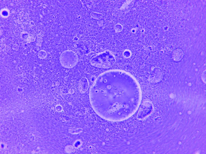
Hematoxylin and eosin staining works by using two dyes that bind to different parts of a cell. Hematoxylin, a basic dye, stains acidic structures like DNA and RNA a blue-purple color. Eosin, an acidic dye, stains basic proteins in the cytoplasm and connective tissue pink. Together, they make tissue structure visible under a microscope.
This combination is called H&E staining. It is the most widely used stain in histology and pathology. Most tissue samples you would see on a pathology slide were prepared this way. Understanding how the two dyes work explains why a slide looks the way it does, and why certain structures stand out in blue or pink.
What Is Hematoxylin And Eosin Staining?
H&E staining is a two-step chemical process that gives colorless tissue visible color. Without it, most tissue under a microscope would appear as a faint, nearly transparent gray.
Hematoxylin is extracted from the heartwood of the logwood tree. On its own, it is not actually a strong dye. It needs a metal ion, called a mordant, to bind tightly to tissue. In routine H&E, the mordant is usually aluminum, in the form of an aluminum salt. The mordant and hematoxylin together form a complex that carries a positive charge. This positive charge is the key to how it stains.
Eosin is a synthetic acidic dye. It carries a negative charge. That opposite charge is why the two dyes stain different things.
The result is a slide where the nucleus of each cell is blue to purple, and the surrounding cytoplasm and connective tissue are shades of pink. The contrast lets a trained eye read cell shape, tissue architecture, and the presence of disease.
How Does Hematoxylin Stain The Nucleus Blue?
Hematoxylin stains the nucleus because DNA and RNA are acidic. They carry a net negative charge. The aluminum-hematoxylin complex carries a positive charge. Opposite charges attract, so the dye complex binds to the nucleic acids inside the nucleus.
This kind of binding is called an electrostatic interaction. The dye is drawn to the negatively charged phosphate groups in the DNA backbone. The nucleus becomes the most densely stained structure on the slide because it holds most of the cell’s nucleic acid.
A detail many people miss: it is the aluminum-hematoxylin complex, not hematoxylin alone, that does the staining. Without the mordant, the dye would not attach well. This is why the mordant is not optional in a working stain.
There are different hematoxylin formulations. Some are designed to stain nuclei more intensely, and some are gentler. The choice affects how crisp the nuclear detail appears, which matters when a pathologist is assessing cell structure.
How Does Eosin Stain The Cytoplasm Pink?
Eosin stains proteins pink because many proteins in the cytoplasm and connective tissue carry a net positive charge. Eosin is negatively charged, so it binds to those positively charged protein regions.
Collagen, the main structural protein in connective tissue, takes up eosin strongly. That is why connective tissue, muscle fibers, and most cell cytoplasm appear pink. Red blood cells also stain bright pink to red because of their high protein content.
Eosin comes in different forms. One common form is slightly more acidic and gives a sharper pink. Another gives a warmer, deeper pink. The difference is subtle but can affect how easily certain structures are seen.
Because eosin stains many proteins, it provides the background that makes the blue nuclei stand out. The two dyes work as a pair. One marks the genetic material, the other marks the protein framework around it.
What Happens To Tissue Before It Is Stained?
Tissue must be prepared before H&E staining can work. The steps matter because each one affects how well the dyes bind.
Fresh tissue is first fixed, usually in a solution of formaldehyde. Fixation cross-links proteins and preserves structure so the tissue does not break down. Without proper fixation, the tissue architecture would be lost and staining would be uneven.
Next, the tissue is processed and embedded in paraffin wax so it can be cut into very thin slices. These slices are typically a few micrometers thick. Then the wax is removed and the tissue is rehydrated through a series of alcohol and water steps, because the dyes work in a watery environment.
Only after these steps does the actual staining happen. The slide is dipped in hematoxylin, then rinsed, then often treated to make the blue color develop properly, then dipped in eosin. Finally it is dehydrated and covered so it can be viewed.
Each step can go wrong. Over-fixation, incomplete wax removal, or poor rinsing can all change the final color and make structures harder to read. This is why quality control in a histology lab matters.
Why Do Some Structures Stain Blue And Others Pink?
The color a structure takes depends on its charge and its composition. Structures rich in nucleic acids, mainly nuclei, take up hematoxylin and look blue to purple. Structures rich in proteins, mainly cytoplasm and connective tissue, take up eosin and look pink.
Here is a quick guide to what typically stains which way:
- Cell nuclei: blue to purple, because of DNA and RNA
- Cytoplasm: pink, because of proteins
- Collagen and connective tissue: pink, because of protein content
- Red blood cells: bright pink to red, because of hemoglobin protein
- Muscle fibers: pink, because of contractile proteins
- Mucus and some other substances: often pale or unstained, because they lack the charged material the dyes bind to
Some structures stain weakly or not at all. Fat, for example, is dissolved away during processing, so it appears as empty spaces. Mucus often looks pale. This is a normal limitation of H&E, not a mistake.
What Can H&E Staining Show And What Can It Miss?
H&E shows tissue architecture and cell structure clearly. A pathologist can see whether cells are normal in shape and arrangement, whether inflammation is present, and whether tissue looks like it should. It is the first stain used for most tissue samples because it gives a broad overview.
What H&E cannot do is identify specific molecules. It shows structure, not chemistry. If a clinician needs to know whether a cell expresses a particular protein, H&E will not answer that. Other techniques, such as special stains or antibody-based methods, are used for that.
H&E also cannot reliably identify every organism or every type of deposit. Some microbes are hard to see on H&E and need special stains. Certain pigments and deposits need other methods to confirm what they are. A pathologist uses H&E as a starting point and adds other tests when needed.
This is an important point about how H&E is used in practice. It is a general-purpose stain that guides the next step. It is rarely the final word on its own for complex questions.
Is H&E Staining Still The Standard Today?
Yes. H&E remains the most widely used stain in diagnostic pathology. It has been in routine use for well over a century, and it is still the default first stain for most tissue specimens.
Newer methods have not replaced it. Techniques like antibody-based staining and molecular testing add information that H&E cannot provide, but they are usually used alongside H&E, not instead of it. The structural overview from H&E tells the pathologist where to look and what to test next.
Part of why H&E persists is practical. It is relatively fast, inexpensive, and well understood. It gives a consistent, readable picture of tissue that most pathologists are trained to interpret. For a first look at a tissue sample, few methods match its combination of speed and clarity.
The dyes and protocols have been refined over the years, but the basic principle, a positive dye for nuclei and a negative dye for proteins, has not changed. That is a sign of how well the method works.
Frequently Asked Questions
Why does hematoxylin stain the nucleus blue?
Hematoxylin binds to the negatively charged DNA and RNA in the nucleus through a positively charged aluminum-dye complex. That electrostatic attraction makes the nucleus take up the blue-purple color.
What is the difference between hematoxylin and eosin?
Hematoxylin is a basic dye that stains acidic structures like nuclei blue, while eosin is an acidic dye that stains basic proteins in cytoplasm and connective tissue pink. They carry opposite charges, which is why they stain different parts of the cell.
Does H&E staining detect cancer?
H&E lets a pathologist see abnormal cell shape and tissue structure, which can suggest cancer, but it does not confirm a diagnosis on its own. Additional tests are often needed to confirm the type and nature of abnormal cells.
Why do some parts of an H&E slide look pale or unstained?
Structures like fat and mucus lack the charged material the dyes bind to, so they appear pale or empty. Fat is usually dissolved during processing, leaving clear spaces on the slide.
