
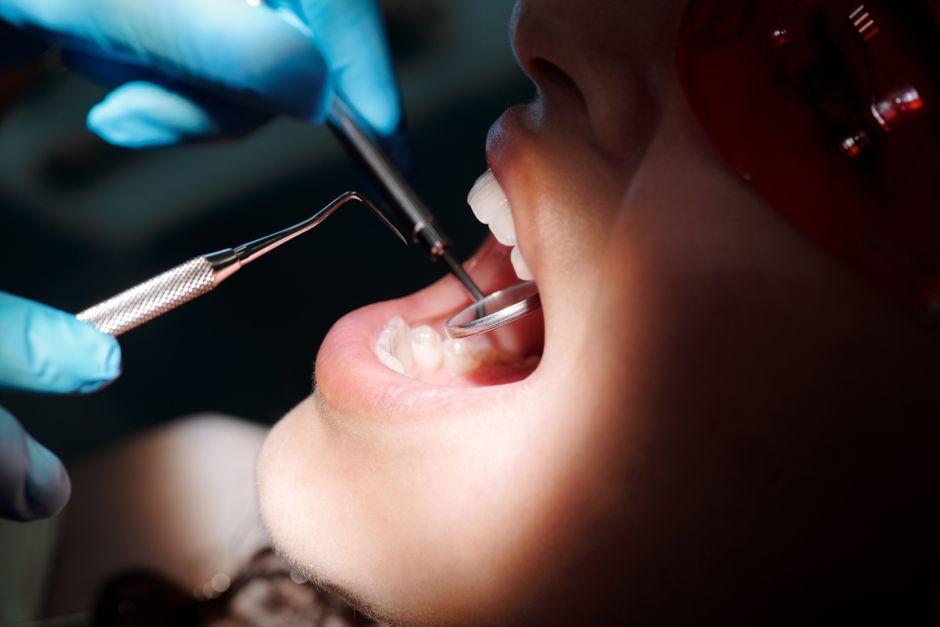
Dentists check for cavities using a combination of visual inspection, tactile probing with a dental explorer, dental radiographs (X-rays), and sometimes additional tools like transillumination or laser fluorescence devices. The most common method remains the visual-tactile exam, where the dentist looks at each tooth surface and feels for soft spots or rough areas with a metal instrument. X-rays are typically used alongside the exam to find decay that isn’t visible to the naked eye, especially between teeth or under existing fillings.
What Happens During a Routine Dental Exam?
A routine dental exam is a systematic process. The dentist or hygienist examines every tooth surface, the gums, and the soft tissues of the mouth. For cavity detection specifically, they focus on the chewing surfaces, the sides of teeth, and the areas near the gumline.
The first step is usually a visual inspection. Good lighting and a small mirror let the dentist see the surfaces of your teeth. They look for discoloration, dark spots, or areas that appear chalky white — a sign that minerals have been lost from enamel. This early stage is called a white spot lesion, and it can sometimes be reversed with fluoride and improved oral hygiene before it becomes a true cavity.
Next comes tactile examination. The dentist uses a dental explorer — a thin metal instrument with a sharp tip — to gently probe the tooth surfaces. A healthy tooth surface feels hard and smooth. Decayed enamel feels soft or sticky when the explorer is pressed against it. This technique has been used for decades and remains a standard part of the exam.
It’s worth noting that probing with a sharp explorer has some limitations. Research has raised concerns that pressing a sharp instrument into an early lesion could potentially break the surface layer and speed up the decay process. Some dental schools and practitioners have moved toward gentler examination techniques, using a blunt probe or relying more on visual and radiographic findings. This is an area where clinical practice varies.
How Do Dental X-Rays Find Cavities?
Dental X-rays, also called radiographs, are one of the most reliable tools for finding cavities that visual examination can miss. They are especially useful for detecting decay between teeth (interproximal decay) and under existing fillings, where a dentist cannot see or feel the problem directly.
There are two main types of X-rays used for cavity detection:
- Bitewing X-rays — These show the crowns of the back teeth and the upper portions of the roots. They are the most common type used for finding cavities between teeth. The dentist or hygienist places a small sensor or film inside your mouth and you bite down on a tab to hold it in place.
- Periapical X-rays — These show the entire tooth from crown to root tip, plus the surrounding bone. They are useful when the dentist suspects decay has reached the pulp or caused an infection at the root.
On an X-ray, a cavity appears as a darker area (called a radiolucency) because decayed tooth structure is less dense than healthy enamel and dentin. The X-ray beam passes through the decayed area more easily, exposing the sensor or film differently than it does through solid tooth structure.
How often you need X-rays depends on your individual risk of decay, your age, and your dental history. The American Dental Association provides guidance on X-ray frequency based on a patient’s risk level, but the decision is ultimately made by your dentist. Adults at low risk may go several years between bitewing X-rays, while those with a history of frequent cavities may need them more often.
What Is Transillumination and How Does It Work?
Transillumination is a technique where a bright light is shone directly through a tooth. Healthy tooth structure scatters the light fairly evenly. Decayed areas, which contain more porous and less mineralized tissue, scatter the light differently and appear as a darker shadow.
This method works best on the front teeth, which are thinner and allow more light to pass through. It is less effective on the back teeth, which are thicker and have more complex surfaces. Some dentists use a fiber-optic light source for transillumination, while others use a device called a DIAGNOdent, which uses laser fluorescence to detect changes in tooth structure.
The DIAGNOdent device works by emitting a specific wavelength of light onto the tooth. Healthy tooth structure reflects very little fluorescence, while decayed areas fluoresce more strongly. The device gives a numerical reading that the dentist can use as one piece of information alongside visual and X-ray findings. It is not a standalone diagnostic tool — it is meant to supplement the standard exam, not replace it.





Why Does the Dentist Sometimes Use a Camera or Digital Scanner?
Many dental offices now use intraoral cameras — small handheld cameras that take magnified digital images of your teeth. These images can be displayed on a screen so you can see what the dentist sees. This is helpful for patient education and for documenting changes over time.
Some offices also use digital impression systems or intraoral scanners, which create a 3D model of your teeth. While these tools are primarily used for making crowns, bridges, and aligners, they can also help the dentist visualize tooth surfaces in detail. They are not typically the primary method for detecting cavities, but they can be an additional aid.
These technologies do not replace the standard visual-tactile exam or X-rays. They add another layer of information that can help the dentist make a more informed decision.
Can Dentists Always See Cavities Right Away?
No. Some cavities are not visible during a routine exam, especially very early ones or those located between teeth. This is why X-rays are important — they can reveal decay that neither the dentist nor the patient can see.
There are also situations where a cavity is visible but its depth is unclear. A dark spot on a tooth could be an active cavity, or it could be a stained area that is not decayed. The dentist uses the explorer, the X-ray, and sometimes additional tools to make that determination.
In some cases, the dentist may recommend a “watch and wait” approach for a suspicious area. This means they will monitor the spot at your next visit to see if it changes. This is more common with early enamel lesions that have not yet broken through the surface. If the area is truly a cavity, it will typically worsen over time without treatment.
What Are the Limitations of Cavity Detection Methods?
No single method is perfect. Visual inspection can miss cavities between teeth or under fillings. X-rays can miss very early decay that hasn’t yet caused enough mineral loss to show up on the image. Transillumination works well on front teeth but is less reliable on back teeth. Laser fluorescence devices can give false positives — meaning they can indicate decay where there is none.
This is why dentists use multiple methods together. The combination of visual inspection, tactile probing, and X-rays gives a much more complete picture than any one technique alone.
It’s also important to understand that the appearance of a cavity on an X-ray or in the mouth does not always mean it needs a filling immediately. The decision to treat depends on whether the decay is active, how deep it is, and whether it has reached the dentin layer. Enamel-only decay that has not broken through the surface may sometimes be managed with fluoride and improved home care rather than drilling.
How Do Dentists Decide If a Cavity Needs a Filling?
The decision to fill a cavity depends on several factors. The dentist considers how deep the decay goes, whether it has reached the dentin (the layer under the enamel), and whether the tooth structure is compromised.
If decay has reached the dentin, a filling is typically recommended. Dentin is softer than enamel and decay spreads through it more quickly. Once decay reaches the pulp — the innermost layer containing nerves and blood vessels — a root canal may be needed instead of a simple filling.
For early enamel decay that has not reached the dentin, some dentists recommend a preventive approach. This might include fluoride varnish, prescription fluoride toothpaste, and more frequent checkups. The goal is to stop the decay process and allow the enamel to remineralize. This approach is more common in patients with good oral hygiene and low cavity risk.
There is some debate in dentistry about when to treat early lesions. Some dentists prefer to fill them early to prevent progression. Others prefer to monitor them and treat only if they worsen. Both approaches have merit, and the choice often depends on the patient’s individual risk factors and the dentist’s clinical judgment.
What Should You Expect at Your Next Dental Visit?
At a typical checkup, you can expect the dentist or hygienist to examine your teeth visually, probe them with an instrument, and possibly take X-rays. They will also check your gums and may use an intraoral camera to show you what they see.
If they find a cavity, they will explain where it is, how deep it is, and what treatment they recommend. If they find an early lesion that doesn’t need treatment yet, they will likely recommend monitoring it and may suggest ways to strengthen your enamel at home.
You can ask questions at any point. If you’re not sure why a particular test or X-ray is being recommended, ask. A good dentist will explain their reasoning and help you understand what they’re looking for.
Frequently Asked Questions
Can dentists find cavities without X-rays?
Yes, dentists can find some cavities through visual inspection and tactile probing alone, especially those on the chewing surfaces or near the gumline. However, X-rays are needed to reliably detect cavities between teeth and under existing fillings that cannot be seen or felt directly.
Are dental X-rays safe for finding cavities?
Dental X-rays use very low levels of radiation, and the risk from a single set of bitewing X-rays is considered very small. The American Dental Association notes that the benefit of detecting decay early generally outweighs the small risk, but X-rays should still be used only when clinically justified.
What does a cavity look like on an X-ray?
On an X-ray, a cavity appears as a darker area because decayed tooth structure is less dense than healthy enamel and dentin. This darker spot is called a radiolucency, and its size and depth help the dentist determine how advanced the decay is.
Can a dentist tell if a cavity is deep just by looking?
Not always. Visual inspection can show that a cavity exists, but it cannot reliably show how deep it goes. X-rays and sometimes additional testing are needed to determine whether decay has reached the dentin or pulp, which affects the type of treatment recommended.
