
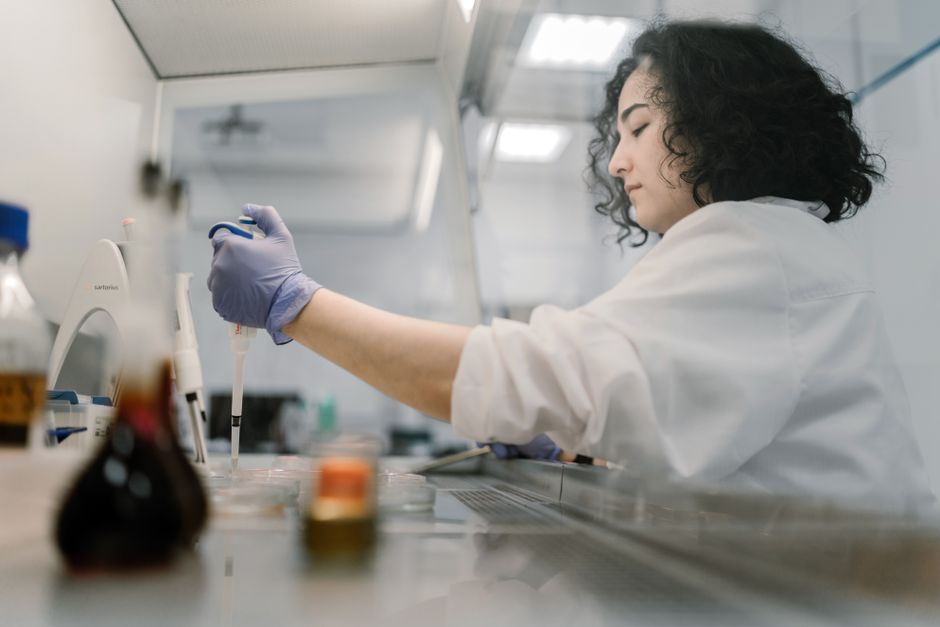
Cell fixation preserves structure by stopping the cell’s own breakdown machinery and locking its molecules in place. Fixatives either cross-link proteins into a stable mesh or rapidly remove water and precipitate molecules into an insoluble network. Either way, the cell is frozen at the moment of fixation, so its shape and internal layout survive the handling, staining, and storage that follow.
That single principle explains why the same tissue can look completely different depending on how it was fixed. A cell is mostly water and enzymes. Left alone, those enzymes digest the cell from within. Fixation halts that process within seconds to minutes and converts a fragile, living structure into something durable enough to section and examine.
What Actually Happens Inside a Cell During Fixation?
Fixation works through two main chemical strategies, and they produce different kinds of preservation.
Cross-linking fixatives, such as formaldehyde, form chemical bridges between proteins. These bridges tie neighboring molecules together into a stable three-dimensional mesh. The cell’s proteins stay roughly where they were, held in place by the new bonds. Because the bonds form throughout the cell, the overall architecture is preserved well.
Precipitating fixatives, such as methanol and ethanol, work differently. They strip water from the cell, which causes proteins and other molecules to clump together and drop out of solution as an insoluble network. This happens fast. The trade-off is that precipitation can distort fine structures more than cross-linking does, because the clumping is less orderly.
In both cases, the enzymes that would normally degrade the cell are themselves fixed. A digestive enzyme cannot chew through a protein it is now bonded to or physically trapped against. That is the core reason fixation protects structure: it disables the cell’s own self-destruction.
Why Does a Cell Need to Be Fixed Before You Can Look at It?
An unfixed cell cannot survive the steps needed to examine it. Microscopy, especially thin sectioning for electron or light microscopy, requires cutting tissue into slices a few micrometers thick or thinner. Living, unfixed tissue is soft, wet, and prone to collapse under a blade.
There is a second problem. Once a cell is removed from its blood supply, its internal environment breaks down. Enzymes called proteases begin digesting proteins, and other processes start dismantling the cell. This is normal biology — the cell’s recycling systems keep running after death. Without fixation, the structure you wanted to observe changes before you can observe it.
Fixation solves both problems at once. It hardens the tissue enough to section cleanly and stops the chemical changes that would blur the picture. The result is a snapshot rather than a moving target.
How Cell Fixation Preserves Structure For Observation
The preservation happens on three levels, and each one matters for what you can see afterward.
Shape and outline. The cell membrane and overall cell contour are held in place. Cross-linking fixatives stabilize the proteins that give the membrane its form, so the cell does not swell, shrink, or rupture during later handling.
Internal organization. Organelles and the cytoskeleton — the internal protein scaffolding — keep their relative positions. This is why a fixed cell still shows where the nucleus sits, how the cytoplasm is arranged, and how structures relate to one another.
Molecular location. Many fixatives anchor molecules where they were, which allows later staining to reveal where specific proteins or other targets are found. This anchoring is central to techniques that detect molecules within intact cells.
It helps to understand what fixation does not do. It preserves location and shape far better than it preserves every fine detail of living function. Some proteins lose their ability to be recognized by certain detection methods after cross-linking, because the chemical bonds alter or hide the sites those methods rely on. This is a well-known limitation, and it is why the choice of fixative is matched to what you want to detect, not chosen at random.
Does the Choice of Fixative Change What You See?
Yes, and often in ways that matter. The fixative is not a neutral step — it shapes the final result.
Formaldehyde-based fixation is widely used because it preserves overall structure well and is compatible with many downstream methods. It cross-links relatively slowly compared with precipitation, so timing and concentration affect the outcome. Methanol and other alcohol-based fixatives act quickly and preserve some molecular targets better, but they can distort fine structure.
There is a practical consequence worth knowing. A structure that appears absent in one preparation may simply have been altered or hidden by the fixative used. Researchers who compare results across studies have to account for this. The same tissue, fixed two different ways, can yield two different pictures — not because the biology changed, but because the preservation method did.
This is why protocols specify fixative type, concentration, and duration together. Changing one variable can shift the result.
What Are the Limits of Fixation?
Fixation is a compromise, not a perfect freeze-frame. It captures structure well but changes chemistry in the process.
Cross-linking introduces new chemical bonds that were not there in the living cell. Those bonds can mask the very sites a later test is designed to detect. Some targets therefore require different fixation or additional steps to become visible again. This is a routine part of laboratory practice, not a rare exception.
Timing also matters. Fixation takes time to penetrate tissue, and during that window the outer layers are preserved before the inner ones. In thicker samples, the center may be less well preserved than the edges. The size of the sample and how quickly the fixative reaches the interior both affect the final quality.
None of this means fixation is unreliable. It means fixation is a tool with known behavior, and interpreting a fixed sample requires knowing how it was prepared. The image you see reflects both the original biology and the preservation method.
Why Does This Matter Outside the Laboratory?
Fixed tissue is the foundation of medical diagnosis. When a biopsy is taken, it is fixed before a pathologist examines it. The diagnosis of many conditions depends on cells that were preserved in a fixed state, then sliced, stained, and viewed under a microscope.
Understanding fixation clarifies something the public rarely considers: a pathology report describes tissue as it appears after chemical preservation, not as it looked in the living body. Skilled pathologists account for this, but the fixed state is always part of what they are reading.
The same principle applies to research. Findings about where molecules sit within cells, or how cells are shaped in disease, rest on fixation having worked correctly. When fixation is done poorly, the conclusions can be wrong.
What Is the Difference Between Fixation and Other Preservation Methods?
Fixation is often confused with freezing, but the two preserve different things.
- Chemical fixation stabilizes structure by cross-linking or precipitating molecules. It is durable and works at room temperature, but it alters chemistry.
- Freezing slows or stops biological processes by lowering temperature. It preserves some molecules in a more native state but can form ice crystals that damage structure if not done carefully.
- Drying removes water and can preserve some features, but it distorts soft tissue severely.
Each method trades one kind of fidelity for another. Chemical fixation favors structural preservation over chemical purity. Freezing favors chemical preservation but risks physical damage. Choosing between them depends on the question being asked.
Frequently Asked Questions
How does cell fixation preserve structure?
It stops the cell’s own enzymes from breaking down proteins and locks molecules in place, either by cross-linking them or by precipitating them into a stable network. This freezes the cell’s shape and internal layout so it survives later handling.
Why is fixation necessary before microscopy?
Unfixed tissue is too soft to slice thinly and begins breaking down once removed from its blood supply. Fixation hardens the tissue and halts that breakdown so the structure stays intact long enough to be examined.
Does fixation change what you can see in a cell?
Yes. Fixatives can alter or hide certain molecular targets, so some proteins are harder to detect after fixation. The method used shapes the final image, which is why protocols specify fixative type and timing.
Is chemical fixation the same as freezing?
No. Chemical fixation stabilizes structure through chemical bonds or precipitation, while freezing slows biological processes through low temperature. Each preserves different features and carries different trade-offs.