
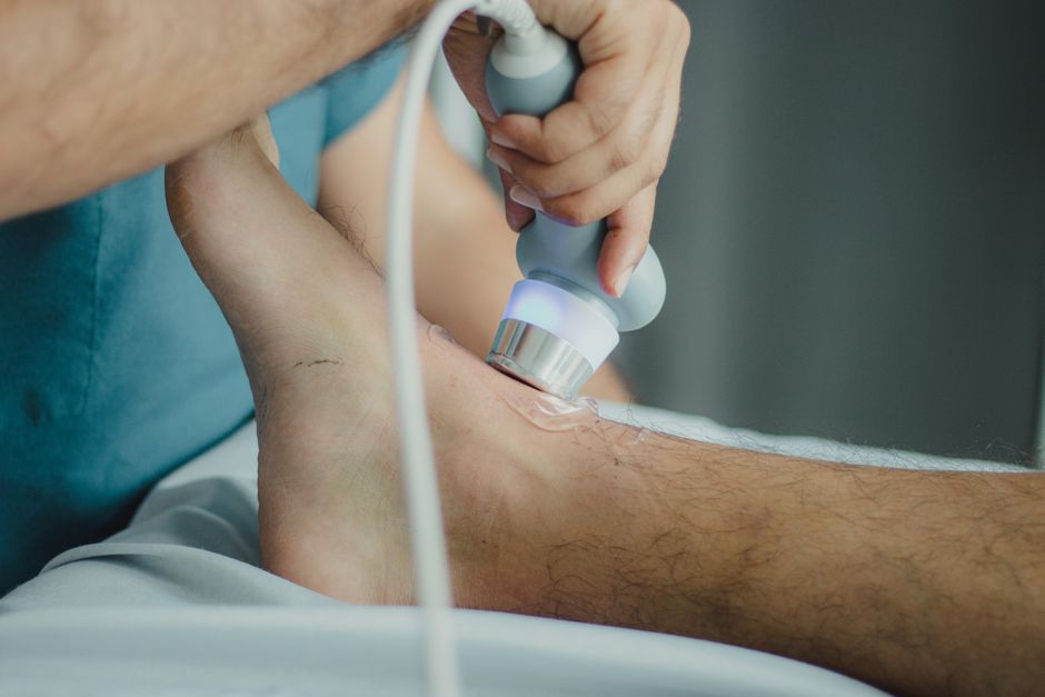
Ultrasound biometric measurements are obtained by using sound waves to create real-time images of a developing baby, then using software tools to trace and measure specific structures like the head, abdomen, and thigh bone. A trained sonographer places a handheld device called a transducer on the mother’s abdomen, which sends high-frequency sound waves into the body. These waves bounce off the baby’s tissues and return to the transducer, where they are converted into a moving image on a screen. The sonographer then freezes the image and uses electronic calipers to mark precise points on the anatomy, allowing the machine to calculate distances in millimeters.
What Exactly Are Biometric Measurements in Ultrasound?
Biometric measurements are the specific numbers that describe a baby’s size at a given point in pregnancy. These are not random estimates. Each measurement follows a strict anatomical protocol so that results are consistent between different sonographers and different machines.
The four most common measurements are the crown-rump length (CRL), biparietal diameter (BPD), head circumference (HC), and femur length (FL). In the first trimester, the CRL is the standard measurement because the baby is curled up and the full length from head to bottom is the most reliable indicator of gestational age. After about 13 weeks, the head and femur measurements become more useful because the baby straightens out and these bones are easier to identify clearly.
The abdominal circumference (AC) is also measured, usually starting in the second trimester. This measurement reflects the size of the liver and the amount of abdominal fat, which makes it a sensitive indicator of fetal growth. Each of these measurements is compared against standard growth charts to determine if the baby is growing at a normal rate.
How Are Ultrasound Biometric Measurements Obtained Step by Step
The process begins with the sonographer applying a water-based gel to the mother’s abdomen. This gel eliminates the air gap between the skin and the transducer, because sound waves travel poorly through air. Without this gel, the image quality would be too poor to obtain reliable measurements.
The sonographer then moves the transducer across the abdomen to find the clearest view of each structure. For the head measurement, the baby’s head must be viewed from a specific angle called the transverse plane. This is a cross-sectional view that shows the head as an oval shape with a visible midline structure in the center. The sonographer adjusts the transducer until the head appears symmetrical, because an angled view will produce falsely large or small numbers.
Once the correct image is frozen on the screen, the sonographer uses the machine’s caliper function to place two markers. For the biparietal diameter, the markers are placed on the outer edge of the near skull bone and the inner edge of the far skull bone. This specific placement is called outer-to-inner and is the standard method used in most clinical settings. The head circumference is obtained by tracing an ellipse around the outside of the skull.
For the femur, the sonographer finds the thigh bone and rotates the transducer until the bone appears as a straight, bright line. Both ends of the bone must be clearly visible. The calipers are placed at the very ends of the bone, excluding the cartilaginous areas that do not appear on ultrasound. The abdominal circumference requires a transverse view of the belly at the level where the umbilical vein enters the liver, and the sonographer traces around the skin line.
Every measurement is taken three times in many practices, and the average is recorded. This reduces the impact of any single difficult image or slight hand movement.
Why Measurement Technique Matters for Accuracy
Small errors in technique can create large errors in clinical decisions. A difference of a few millimeters in the head measurement can change the estimated gestational age by several days. In the second trimester, this can shift a woman’s due date. In the third trimester, it can change whether a baby is classified as growing normally or as small for gestational age.
The most common source of error is an incorrect imaging plane. If the head is measured from an angle rather than a true cross-section, the diameter will appear smaller than it actually is. If the abdomen is measured too high or too low, the circumference may include the ribs or miss the liver, both of which distort the result.
Another source of error is the caliper placement itself. Different practitioners may place markers slightly differently on the same image. This is why professional guidelines emphasize standardized techniques. The International Society of Ultrasound in Obstetrics and Gynecology publishes detailed protocols that describe exactly which landmarks to use and where to place the calipers. Following these protocols keeps measurements comparable across different clinics and different countries.
Fetal position also affects accuracy. If the baby is moving actively, the sonographer may need to wait for a moment of stillness. If the baby is positioned with its spine directly facing the transducer, the sound waves may not penetrate clearly, and the sonographer may ask the mother to change position or return for a repeat scan later.




How Measurements Are Used to Estimate Gestational Age
The crown-rump length is the most accurate measurement for dating a pregnancy. In the first trimester, between about 7 and 13 weeks, the CRL can estimate gestational age within about 5 to 7 days. This is because all babies grow at nearly the same rate early in pregnancy, regardless of their eventual birth weight.
After the first trimester, growth rates become more variable. Some babies are genetically larger or smaller. This means that head and femur measurements become less reliable for dating. In the second trimester, the accuracy drops to about 7 to 10 days. In the third trimester, the margin of error can be 2 to 3 weeks or more.
For this reason, the due date is set based on the earliest reliable ultrasound, ideally the first-trimester CRL. Later scans are not used to change the due date unless the discrepancy is very large. Instead, later measurements are used to track growth over time.
Fetal weight estimation uses a formula that combines several measurements, typically the head circumference, abdominal circumference, and femur length. These formulas are statistical models based on large populations of babies with known birth weights. They provide an estimate, not an exact number, and the margin of error is roughly 10 to 15 percent.
What Can Affect the Quality of Measurements
Several factors can make measurements harder to obtain. Maternal body habitus is one of them. Sound waves must travel through the abdominal wall, and thicker tissue weakens the signal. This can produce a grainy image where the edges of bones are less distinct, making caliper placement more difficult.
The amount of amniotic fluid matters as well. Sound waves travel well through fluid, so adequate amniotic fluid provides a clear window to the baby. If the fluid level is low, the baby may be pressed against the uterine wall, and the image may be less clear.
The position of the placenta can also interfere. If the placenta is located on the front wall of the uterus, directly beneath the transducer, it can scatter the sound waves. This condition is called an anterior placenta, and it often requires the sonographer to angle the transducer differently or scan from the side.
Finally, the baby’s own position is a major factor. A baby facing away from the transducer may hide the facial features, but the head and femur measurements are usually still obtainable. A baby in a breech position can make the head measurement more difficult because the head is tucked under the mother’s ribs, which can shadow the image.
When image quality is poor, the sonographer will typically state that the measurement has limited reliability. Some clinics will offer a repeat scan on another day rather than report a number that may be inaccurate.
Is There Any Risk or Discomfort Involved
Diagnostic ultrasound has been used in pregnancy for decades, and no harmful effects have been confirmed when it is used appropriately by trained professionals. The sound waves used in standard obstetric ultrasound are at a low power level, and the examination is typically completed within 20 to 30 minutes.
The procedure itself is not painful. The gel may feel cold, and the sonographer may need to press firmly to obtain a good image, particularly in later pregnancy. Some mothers feel mild discomfort if the transducer presses on a full bladder, which is sometimes required in early pregnancy to improve the view of the uterus.
It is important to note that ultrasound is a diagnostic tool, not a form of treatment. It does not expose the mother or baby to ionizing radiation, which is the type of radiation used in X-rays. However, the American College of Obstetricians and Gynecologists and other professional bodies recommend that ultrasound be used only when there is a medical reason, not for keepsake images or non-medical purposes.
How Do Measurements Compare to Growth Charts
After each measurement is obtained, it is plotted on a growth chart. These charts show the range of normal measurements for each week of pregnancy. A baby at the 50th percentile is exactly average for that gestational age. A baby at the 10th percentile is smaller than 90 percent of babies at that age, and a baby at the 90th percentile is larger than 90 percent of babies.
The most commonly used charts in the United States come from the Hadlock formula, which was developed from a large sample of pregnancies in the 1980s and 1990s. More recent charts, such as the INTERGROWTH-21st standards, were developed from an international population of healthy mothers with optimal nutrition and healthcare. The choice of chart can affect whether a baby is classified as growth-restricted, so many clinics now specify which chart they use in their reports.
Measurements are also compared over time. A single measurement gives a snapshot, but the trend across multiple scans gives a more complete picture. If a baby’s abdominal circumference drops from the 40th percentile at 28 weeks to the 5th percentile at 34 weeks, that pattern is more concerning than a single low measurement at 34 weeks.
Frequently Asked Questions
Do ultrasound biometric measurements hurt the baby?
No evidence shows that diagnostic ultrasound harms a developing baby when used appropriately by trained professionals. The sound waves are at low power, and the scan typically lasts less than 30 minutes.
How accurate are ultrasound measurements for due date?
First-trimester crown-rump length measurements estimate the due date within about 5 to 7 days. Later measurements are less accurate for dating because fetal growth rates become more variable after the first trimester.
Why do two different ultrasounds give different measurements?
Fetal position, maternal tissue, and the exact angle of the transducer can all affect measurements. Small differences in caliper placement between sonographers also contribute to variability.
Can ultrasound measurements predict the baby’s birth weight exactly?
No, ultrasound weight estimates have a margin of error of roughly 10 to 15 percent. They are useful for identifying growth trends but are not exact predictions of birth weight.
