
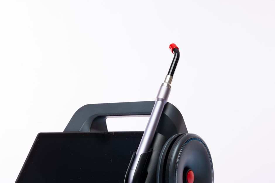
A portable fundus camera is a handheld device that takes detailed pictures of the back of the eye, including the retina, optic nerve, and blood vessels. It works by shining light through the pupil and capturing the reflection with a built-in digital sensor. These devices allow eye doctors and primary care providers to examine the retina outside of a traditional clinic setting, making eye screening more accessible.
What Is a Fundus Camera and What Does It Capture?
The term “fundus” refers to the inside back surface of the eye. This area includes the retina, which detects light, and the optic nerve, which carries visual information to the brain. A fundus camera is essentially a specialized microscope with a camera attached.
The images it captures reveal the health of small blood vessels in the retina. These vessels are often affected by systemic conditions before symptoms appear. For this reason, retinal imaging is useful for detecting signs of diabetic retinopathy, glaucoma, and age-related macular degeneration.
Portable versions of this technology are smaller than traditional tabletop models. They are designed to be held by a clinician or mounted on a small stand. The goal is the same as a clinic-based camera, but the portability allows for use in community health centers, nursing homes, and remote locations.
How Does the Camera Actually Take a Picture?
The process seems simple but involves precise optics. The camera uses a low-intensity flash of light that travels through the pupil and illuminates the retina. Light reflects off the retinal tissue and travels back through the pupil into the camera’s sensor.
The key component is the optical system. The lens must focus light through the cornea and lens of the patient’s eye, then onto the retina, and capture the returning light on a digital sensor. This requires a specific alignment between the camera, the pupil, and the retina.
Most portable fundus cameras use a technology called non-mydriatic imaging. This means they do not require eye drops to dilate the pupil. The camera uses infrared light to focus and align before capturing the image with a visible flash. This makes the process faster and more comfortable for patients.
The entire capture takes seconds. The digital sensor records the image, which can be viewed immediately on a screen. The clinician can assess image quality on the spot and retake the photo if needed.
What Makes a Portable Fundus Camera Different From a Traditional One?
Traditional fundus cameras are large, fixed instruments. They require patients to sit upright with their chin and forehead pressed against a support. These systems produce high-resolution images and are the standard in hospital ophthalmology departments.
Portable models sacrifice some image quality for convenience and accessibility. They are lighter, more compact, and can be used on patients who cannot sit upright or travel to a specialist. This is especially important for elderly patients, people with mobility limitations, and those in rural areas.
Another difference is the field of view. Traditional cameras can capture a wider area of the retina in a single image. Portable cameras often capture a narrower field, meaning the clinician may need to take multiple images to cover the same area.
However, portable models have improved significantly in recent years. Many now produce images sufficient for screening and monitoring chronic conditions. The trade-off between portability and image quality continues to narrow as technology advances.





Who Uses Portable Fundus Cameras and Why?
Ophthalmologists and optometrists are the primary users. But portable cameras have expanded the range of providers who can perform retinal screening. Primary care physicians, endocrinologists, and even trained technicians can use these devices with proper training.
The most common use is screening for diabetic retinopathy. Diabetes affects the small blood vessels in the retina, and early detection allows for timely treatment. Regular screening is recommended for people with diabetes, but many do not see an eye specialist annually. Portable cameras allow screening during routine primary care visits.
Other uses include monitoring glaucoma, assessing the optic nerve, and evaluating patients with high blood pressure. The retina is one of the few places in the body where blood vessels can be viewed directly without surgery. This makes it a valuable window into cardiovascular health.
Portable cameras are also used in emergency departments and urgent care settings. They help evaluate patients with sudden vision changes, headaches, or suspected increased pressure inside the skull.
How Accurate Are Portable Fundus Cameras Compared to Traditional Imaging?
Research generally shows that portable fundus cameras are reliable for detecting major retinal conditions. Studies comparing portable and traditional cameras have found similar rates of detecting diabetic retinopathy and other common eye diseases.
Image quality varies by model and operator skill. A poorly focused image can miss subtle findings. But for screening purposes, where the goal is to identify who needs a full specialist examination, portable cameras perform well.
Some research suggests that image quality is lower in patients with cataracts or small pupils. These conditions make it harder for light to reach the retina and return to the sensor. In these cases, the clinician may need to refer the patient for traditional imaging with pupil dilation.
The accuracy also depends on the reader. Some programs use trained graders who review images remotely. Others use automated software that flags suspicious findings. Both approaches have been shown to be effective when implemented properly.
What Are the Limitations of Portable Fundus Cameras?
Portable cameras have real limitations that patients and providers should understand. They are not a replacement for a comprehensive eye examination by an ophthalmologist.
The most significant limitation is the field of view. A narrow field means peripheral retinal lesions may be missed. Conditions that start at the edges of the retina, such as retinal tears or detachments, may not appear in the captured image.
Another limitation is the inability to perform certain tests. A fundus camera takes a photograph, but it does not measure visual acuity, test peripheral vision, or evaluate how the eye focuses light. These tests require different equipment and are essential for a complete eye examination.
Patient cooperation is also important. The patient must keep their eye open and steady for a few seconds. Blinking or movement can blur the image. While most patients can comply, some may find the flash uncomfortable.
Cost is another factor. While portable cameras are less expensive than traditional systems, they still represent a significant investment for smaller clinics. The cost may be justified by the ability to screen more patients and reduce referrals to specialists.
How Do You Prepare for a Retinal Imaging Session?
In most cases, no preparation is needed. The patient sits in a chair or lies down, and the clinician positions the camera in front of the eye. The patient is asked to look at a fixed point while the camera captures the image.
Patients who wear glasses or contact lenses may be asked to remove them. This allows the camera to focus directly on the eye. The clinician may use a small lens to compensate for refractive errors in the patient’s vision.
The procedure is painless and takes only a few minutes per eye. The flash can be bright but is brief. Some patients see a temporary afterimage, which fades within seconds or minutes.
No clinical guidelines currently require fasting, special medication adjustments, or other preparation for fundus photography. Patients can eat, drink, and take their regular medications as usual before the procedure.
What Happens After the Images Are Taken?
The images are stored digitally and can be reviewed immediately. The clinician looks for abnormalities such as bleeding, swelling, or changes in the optic nerve. If the images are clear and normal, the patient may be told no follow-up is needed.
If the images show concerning findings, the patient is referred to an eye specialist for a more detailed examination. The referral is not a diagnosis. It means the images suggest something that needs further evaluation.
Some portable camera systems include software that analyzes images automatically. This software can flag potential signs of diabetic retinopathy or other conditions. However, automated analysis is not a substitute for a trained clinician’s review.
The images become part of the patient’s medical record. They can be compared with future images to track changes over time. This is particularly valuable for monitoring chronic conditions like diabetes or glaucoma.
Frequently Asked Questions
Is a portable fundus camera painful?
No, the procedure is painless. The patient may see a bright flash, and some people experience a brief afterimage that fades quickly.
Do I need eye drops to dilate my pupils for a portable fundus camera?
Most portable cameras use non-mydriatic technology, meaning they do not require dilating drops. Some models can also capture images with dilated pupils for higher detail when needed.
Can a portable fundus camera detect glaucoma?
Yes, it can capture images of the optic nerve, which is the part of the eye affected by glaucoma. However, it cannot measure eye pressure or test peripheral vision, so it is not a complete glaucoma examination.
How long does a retinal imaging session take?
The imaging itself takes only a few minutes for both eyes. The entire visit, including positioning and review of the images, typically takes less than 15 minutes.
