
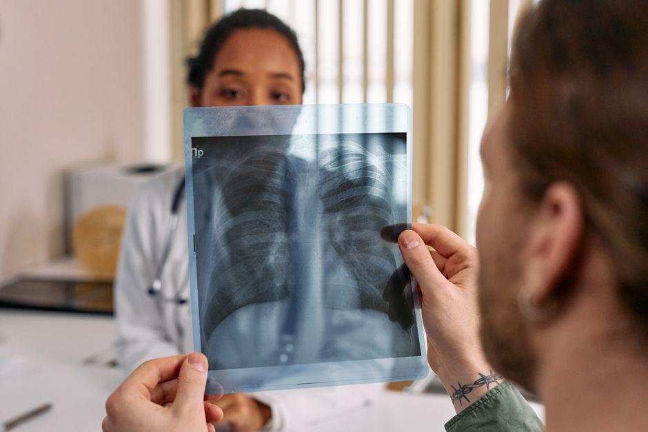
A PET scan can light up a benign lung nodule, but most benign nodules do not appear bright on PET imaging. The test measures how much glucose-based tracer a spot absorbs, and some non-cancerous conditions — especially active infections and inflammation — absorb enough to look “hot.” That overlap is why PET results are always read alongside the size of the nodule, its appearance on CT, and the person’s history.
The short version: PET is not a cancer test with a yes-or-no answer. It is one piece of a larger picture, and its usefulness depends heavily on how big the nodule is and what else is going on in the lung.
How Does a PET Scan Actually Detect Activity in a Lung Nodule?
Most PET scans for lung nodules use a radioactive form of glucose called FDG, short for fluorodeoxyglucose. Cells that are burning through a lot of energy pull in more of it than quiet cells do. The scanner then shows where that tracer collected.
Cancer cells tend to be metabolically busy, so they often show up as bright spots. But so do many other cells. White blood cells fighting an infection, immune cells responding to inflammation, and healing tissue all consume glucose at high rates. The tracer does not know the difference between a tumor and a lymph node doing its job.
This is the core problem with PET for lung nodules. It measures metabolic activity, not identity. A bright spot means “something here is using a lot of energy.” It does not mean “this is cancer.”
There is also a size issue that matters more than most people realize. PET scanners have limited resolution. Nodules smaller than roughly 8 to 10 millimeters often do not show up clearly at all, whether they are benign or malignant. A “negative” PET on a tiny nodule is not reassuring — it may simply mean the nodule was too small to see.
Do Benign Lung Nodules Light Up On A PET Scan?
Yes, some do. This is not a rare technicality. It is a well-documented limitation of the test.
Certain benign conditions are known to produce false-positive results on lung PET scans:
- Infections — active pneumonia, tuberculosis, fungal infections, and lung abscesses all draw in immune cells that consume glucose
- Inflammation — sarcoidosis, rheumatoid lung disease, and other inflammatory conditions can light up
- Post-surgical or post-radiation changes — healing tissue is metabolically active for weeks to months
- Benign tumors — some hamartomas and other non-cancerous growths can absorb tracer
The reverse is also true. Some cancers, particularly slow-growing ones like certain carcinoid tumors and some adenocarcinomas, do not light up brightly. A nodule that looks quiet on PET is not automatically safe.
So PET produces both false positives and false negatives. Its value comes from how it shifts the odds when combined with other information, not from any single result.
Why Does Inflammation Look Like Cancer on a PET Scan?
Because inflammation and cancer share a feature: both involve cells that are working hard.
When tissue is inflamed, the body sends in macrophages and other immune cells. These cells are highly active. They need energy, and they get it by taking up glucose. FDG is a glucose analog, so it gets pulled in too. On the scan, that patch of inflammation glows.
Infection works the same way. A pocket of bacteria triggers an immune response, and that response is metabolically expensive. The result is a bright spot that has nothing to do with cancer.
This is not a flaw in the scanner. It is a fundamental limitation of using metabolic activity as a proxy for cancer. The test is doing exactly what it was designed to do — it just cannot tell you why the cells are busy.
What Does the Standardized Uptake Value (SUV) Tell You?
PET reports often include a number called the SUV, or standardized uptake value. It is a rough measure of how much tracer a spot absorbed. Higher numbers generally mean more metabolic activity.
There is no single SUV cutoff that cleanly separates benign from malignant. Higher values raise concern; lower values lower it. But the overlap is substantial. Some infections produce very high SUVs. Some cancers produce low ones.
Clinicians use SUV as one input among several. They also look at:
- The size and shape of the nodule on CT
- Whether it has grown over time on repeat scans
- Its edges — smooth, irregular, or spiculated
- Whether there is calcification, which often suggests a benign cause
- The person’s age, smoking history, and exposure history
A single number rarely decides anything. The pattern across all these features does.
When Is a PET Scan Actually Useful for a Lung Nodule?
PET is most helpful for nodules that are large enough to be seen clearly and that fall into an uncertain middle ground on CT. In that setting, it can shift the probability up or down and help guide whether to biopsy, watch, or proceed to surgery.
It is less useful — sometimes not useful at all — for:
- Very small nodules, where the scanner cannot resolve them reliably
- Nodules that already look clearly benign on CT, such as those with certain calcification patterns
- Nodules that already look highly suspicious, where the result would not change the plan
Guidelines from professional radiology and chest medicine groups generally do not recommend PET for routine evaluation of small nodules. The test has a specific role, and using it outside that role tends to produce confusing results.
What Happens If a Benign Nodule Lights Up?
This is the scenario that worries people most, and it is worth walking through honestly.
A bright nodule on PET raises the concern for cancer. That does not mean cancer is present. It means the next step is usually to get a tissue sample — a biopsy — or to follow the nodule closely with repeat imaging.
If a biopsy shows benign tissue, the case is usually closed, though follow-up imaging may still be done depending on the specific finding. If the nodule is in a spot that is hard to biopsy, doctors may recommend a period of watchful waiting with repeat CT scans to see whether it changes.
The frustration here is real. A false-positive PET can lead to a biopsy that turns out to be unnecessary. This is a known trade-off of the test. It is also why PET is not used as a blanket screening tool for lung nodules.
Does a Negative PET Scan Mean a Nodule Is Benign?
No. A negative PET — one where the nodule does not light up — lowers the probability of cancer, but it does not rule it out.
Small nodules may not appear on PET regardless of what they are. Slow-growing cancers may not consume enough glucose to show up. And some tumor types are simply not FDG-avid, meaning they do not take up the tracer well.
For these reasons, a negative PET is usually combined with continued surveillance rather than treated as a final answer. The nodule still gets watched.
The Bottom Line on PET and Benign Nodules
PET scans detect metabolic activity, and metabolic activity is not the same as cancer. Benign nodules can light up, especially when infection or inflammation is involved. Malignant nodules can stay dark, especially when they are small or slow-growing.
The test is useful in the right context and misleading outside it. If you have a lung nodule and are trying to understand what a PET result means, the most useful question is not “did it light up?” but “what does this result change about the plan?” That is the question your care team is actually answering.
Frequently Asked Questions
Can a benign lung nodule show up bright on a PET scan?
Yes. Infections, inflammation, and some benign tumors can absorb enough tracer to appear bright. This is a well-known cause of false-positive results.
What size nodule is too small for a PET scan to evaluate?
Nodules smaller than roughly 8 to 10 millimeters often cannot be reliably assessed on PET. A negative result on a nodule that small does not rule out cancer.
Does a PET scan tell you for sure whether a lung nodule is cancer?
No. PET measures metabolic activity, not tissue type, so it cannot confirm or exclude cancer on its own. A biopsy is needed for a definitive answer.
If a PET scan is negative, does that mean the nodule is harmless?
Not necessarily. A negative PET lowers the concern for cancer but does not eliminate it, which is why follow-up imaging is often still recommended.
