
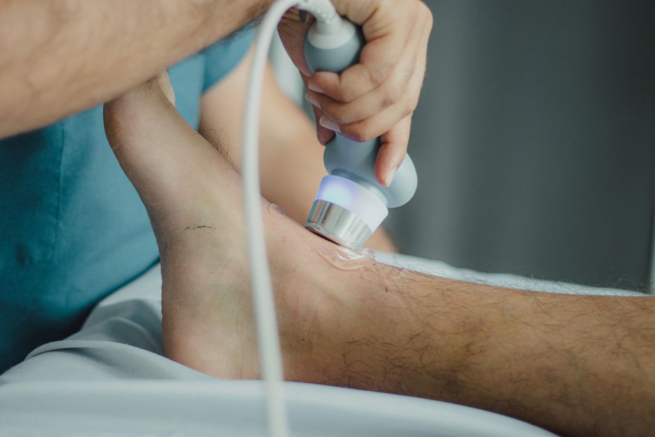
Yes, a lipoma can usually be seen on an ultrasound. A lipoma is a benign growth of fat cells that sits under the skin, and ultrasound uses sound waves to create a picture of that tissue. On the screen, a typical lipoma appears as a soft, oval or rounded mass just beneath the skin, and it tends to look lighter or similar in shade to the surrounding fat.
Ultrasound is often the first imaging test a doctor orders for a lump that feels soft and movable. It is quick, painless, uses no radiation, and can usually confirm whether a lump is made of fat. That said, ultrasound has limits, and some lipomas need a different test to be sorted out from other lumps.
What Does a Lipoma Look Like on an Ultrasound?
A lipoma has a fairly recognizable signature on ultrasound. Doctors describe it as a well-defined mass that sits in the layer of fat just under the skin. It usually looks similar to or slightly different from the fat around it, which is one reason it can blend in.
Several features help confirm it is fat rather than something else:
- It is usually oval or rounded, with smooth, clear edges.
- It sits within the fatty layer, not deep inside muscle.
- It tends to be soft when pressed with the ultrasound probe.
- It usually has few or no blood vessels running through it.
- It often has thin, faint bands running through it.
That last point matters. Fat cells are separated by thin walls of connective tissue, and those walls can show up as faint stripes. This pattern is common in lipomas and helps distinguish them from other soft lumps.
One useful detail: a lipoma is typically avascular, meaning it has very little blood flow. A doctor can check this with a setting called color Doppler, which shows blood movement. A lump with a lot of blood flow is less likely to be a simple lipoma and may need a closer look.
Can You See a Lipoma on an Ultrasound? How Reliable Is It?
Ultrasound is quite good at identifying a typical lipoma, but it is not perfect. For a soft, movable lump under the skin, it is often the first and sometimes the only test needed. Many doctors can make a confident call based on the ultrasound picture combined with how the lump feels.
Reliability depends on a few things:
- Where the lump is. Lipomas near the surface are easier to see clearly than deep ones.
- The size of the lump. Very small or very large lumps can be harder to assess.
- The skill and equipment of the person scanning. A trained operator with a good machine gets better images.
- Whether the lump is a typical lipoma or a variant. Some fatty lumps have unusual features that blur the picture.
When the ultrasound shows a classic fatty mass and the lump feels typical, most clinicians are comfortable calling it a lipoma. When the picture is unclear or unusual, they may order more imaging or a tissue sample.
There is an honest limit here. Ultrasound shows the shape and texture of a lump. It cannot always tell you with certainty what the cells are. That is why unusual features sometimes prompt further testing.
What Else Can Look Like a Lipoma on Ultrasound?
Several other lumps can mimic a lipoma, and this is one of the real reasons ultrasound matters. Not every soft lump is harmless fat.
Other possibilities include:
- Liposarcoma. A rare cancer of fat tissue. It can look similar to a lipoma but often has features that raise concern, such as deeper location, irregular edges, or more blood flow.
- Epidermoid cyst. A fluid- or debris-filled sac that often sits closer to the skin surface.
- Fibroma or other fibrous lumps. These are made of connective tissue, not fat, and look different on ultrasound.
- Enlarged lymph nodes. These can appear as lumps under the skin in certain areas.
- Hematoma or fluid collection. A pocket of blood or fluid after an injury.
This is why a doctor does not rely on the ultrasound alone. The feel of the lump, its location, how long it has been there, and whether it is changing all factor into the assessment.
Certain warning signs make a lump more concerning and more likely to need a biopsy:
- It is growing quickly.
- It feels firm or fixed in place rather than soft and movable.
- It is deep rather than just under the skin.
- It is painful or causes other symptoms.
- The ultrasound shows irregular edges or a lot of blood flow.
None of these signs prove a lump is cancer. They simply mean a closer look is warranted.





How Does Ultrasound Compare to Other Imaging Tests for Lipomas?
Ultrasound is not the only way to look at a fatty lump. Each imaging tool has strengths, and doctors choose based on the situation.
| Test | What it shows | Best for |
|---|---|---|
| Ultrasound | Shape, texture, depth, and blood flow of a lump | Soft lumps just under the skin |
| MRI | Detailed pictures of soft tissue, including deep lumps | Deep, large, or unclear lumps |
| CT scan | Cross-sectional images, including bone and deep tissue | Certain deep or complex cases |
| Biopsy | A sample of the tissue itself | Lumps that look unusual or concerning |
For a typical lump under the skin, ultrasound is usually enough. MRI gives a more detailed view and is often used when a lump is deep, large, or does not look like a simple lipoma. A biopsy removes a small sample of tissue so it can be examined under a microscope. That is the only way to be fully certain what the cells are.
It helps to understand the trade-offs. Ultrasound is fast, cheap, and has no radiation. MRI gives more detail but costs more and takes longer. A biopsy gives a definite answer but is a minor procedure. Doctors weigh these factors against how suspicious the lump appears.
When Should You Get a Lump Checked?
Any new lump deserves a look, especially if it is growing, firm, painful, or deep. Most lumps turn out to be harmless, but the only way to know is to have a clinician examine it.
See a doctor if a lump:
- Is getting bigger over weeks or months.
- Feels hard or stuck in place.
- Is painful or tender.
- Sits deep under the skin or in muscle.
- Comes with fever, weight loss, or night sweats.
A soft, movable lump that has been stable for years is less concerning, but it is still reasonable to have it checked once. A clinician can often tell a lot just by feeling the lump and looking at the skin over it.
Here is a point worth making clearly: size alone does not tell you whether a lump is dangerous. Some large lipomas are harmless, and some small lumps are not. What matters more is how the lump behaves over time and what the imaging shows.
What Happens If Ultrasound Is Not Enough?
Sometimes the ultrasound does not give a clear answer. In that case, a doctor may recommend more steps.
If a lump looks unusual, the next step is often an MRI. This gives a more detailed view of the tissue and can help show whether a lump has features that suggest it is not a simple lipoma. For deep lumps, MRI is frequently the preferred test.
If imaging still leaves doubt, or if the lump has concerning features, a biopsy may be recommended. A biopsy takes a small sample of the tissue so a pathologist can examine the cells directly. This is the most reliable way to confirm what a lump is.
It is worth being honest about the uncertainty here. Imaging can strongly suggest a lipoma, but it cannot always rule out every other possibility with total certainty. That is not a flaw in ultrasound. It is a limit of what any imaging test can do without looking at the tissue itself.
Most people with a confirmed lipoma need no treatment at all. A lipoma is benign, and many stay the same size for years. Removal is usually optional and is done mainly if the lump is painful, growing, or bothersome. A doctor can discuss whether watching it or removing it makes more sense in a given case.
Frequently Asked Questions
Can a lipoma be diagnosed with ultrasound alone?
Often yes, especially for a soft lump just under the skin that has the typical features of fat on the scan. When the picture is unclear or the lump looks unusual, a doctor may order an MRI or a biopsy for a more definite answer.
What does a lipoma look like on an ultrasound?
It usually appears as a smooth, oval or rounded mass in the fatty layer under the skin, with clear edges and little blood flow. It often looks similar in shade to the fat around it, sometimes with faint thin bands running through it.
Can ultrasound tell a lipoma from cancer?
Ultrasound can raise or lower suspicion based on features like edges, depth, and blood flow, but it cannot fully rule out cancer on its own. A biopsy is the only way to confirm what the cells actually are.
Do lipomas need to be removed?
Most lipomas do not need removal because they are benign and often stay stable for years. Removal is usually optional and is considered mainly if the lump is painful, growing, or bothersome.
