
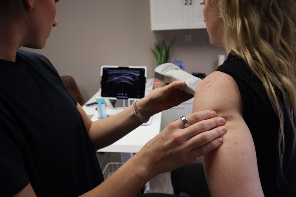
Yes, ultrasound can show tendon damage and tears, and it is one of the first imaging tools doctors use to evaluate these injuries. A skilled technician can see the internal structure of a tendon in real time, identifying inflammation, partial tears, and complete ruptures. It is a safe, quick, and dynamic test that allows the doctor to move the joint while watching the tendon on screen, which often reveals problems that static images miss.
How Does Ultrasound Detect Tendon Problems?
Ultrasound works by sending high-frequency sound waves into the body. These waves bounce off tissues and return to the transducer, creating a live image on a monitor. Healthy tendons appear as a bright, organized, fiber-like pattern. When a tendon is damaged, that pattern changes.
Tendon injuries typically fall into two categories: tendinopathy and tears. Tendinopathy is chronic damage from overuse. The tendon thickens, develops areas of low signal, and may have increased blood flow. A tear is an actual break in the tendon fibers. Ultrasound shows both clearly, but they look different on the screen.
For a partial tear, the image shows a dark gap within the tendon that does not go all the way through. The tendon fibers appear disrupted or thinned in one area. A full-thickness tear shows a complete break where the tendon separates into two pieces. Often, the gap fills with fluid or scar tissue, making it even easier to see.
What Can Ultrasound See That Other Tests Cannot?
The main advantage of ultrasound is that it is dynamic. MRI takes a series of static pictures while you lie still. Ultrasound lets the examiner move your joint through its full range of motion while watching the tendon in real time. Some tears only separate when the tendon is under tension. A tendon that looks intact at rest may open up like a gap when you flex the muscle, revealing a tear that an MRI could miss.
Ultrasound also allows for direct comparison. The examiner can scan the injured side and then the healthy side using the same settings. This side-by-side comparison helps identify subtle changes in thickness, texture, or movement that might otherwise be overlooked.
Another practical advantage is cost and speed. An ultrasound is generally less expensive than an MRI and takes about 15 to 30 minutes. There is no radiation exposure and no need to lie still inside a closed tube, which makes it more comfortable for people who are claustrophobic or in significant pain.
Which Tendons Are Best Evaluated by Ultrasound?
Ultrasound works best on tendons that are close to the skin surface. These are the tendons doctors scan most often:
- Rotator cuff tendons in the shoulder — the most common tendon ultrasound
- Achilles tendon at the back of the ankle
- Patellar tendon below the kneecap
- Common extensor tendon at the elbow, often called tennis elbow
- Peroneal and tibialis tendons around the ankle
- Hand and wrist flexor tendons
Deeper tendons are harder to see with ultrasound. The tendons deep inside the hip or behind the shoulder joint are better evaluated with MRI because the sound waves lose strength as they travel through muscle and bone. For most superficial tendons, however, ultrasound is highly accurate.
Research consistently shows that ultrasound is comparable to MRI for detecting full-thickness rotator cuff tears. Some studies indicate that ultrasound may be slightly less reliable for identifying small partial tears in certain locations, but operator skill plays a major role in accuracy. An experienced examiner who performs many tendon scans each week will get better results than someone who rarely does them.





What Are the Limitations of Ultrasound for Tendons?
Ultrasound is operator-dependent. The quality of the exam relies heavily on the skill and experience of the person performing it. A less experienced technician can miss subtle tears or mistake scar tissue for a healthy tendon. This is different from an MRI, where the machine does most of the work and a radiologist interprets the images afterward.
Ultrasound also has a limited field of view. The transducer only captures a small area at a time. A large tear or a complex injury involving multiple structures may require many separate scans to map out fully. In these cases, an MRI provides a broader overview of the entire joint in a single test.
Bone blocks sound waves completely. Areas where a tendon passes behind a bony prominence may be impossible to see fully. The examiner must angle the transducer around the bone, and sometimes that is not enough to get a complete picture of the tendon.
Finally, ultrasound cannot see inside the bone itself. If the doctor suspects a fracture or bone bruise in addition to a tendon injury, an X-ray or MRI would be needed to evaluate the bone.
How Accurate Is Ultrasound Compared to MRI?
For full-thickness tendon tears, ultrasound and MRI have similar accuracy. Both are excellent at identifying a complete rupture. For partial tears, the picture is more complicated. Some research suggests MRI detects a higher percentage of partial tears, particularly in the shoulder. Other studies show that when performed by experienced operators, ultrasound matches MRI for partial tears as well.
The practical reality is that both tests have their place. A doctor may start with ultrasound because it is fast, inexpensive, and can be done in the office. If the ultrasound is inconclusive or the injury seems more complex, an MRI provides a more detailed look at the surrounding tissues, including cartilage, ligaments, and bone marrow.
Ultrasound does have one clear advantage over MRI for guiding treatment. Because it is real-time, doctors can use it to guide injections precisely into the damaged area. This is not possible with MRI. The live image ensures the needle goes exactly where it needs to go, which improves the accuracy of corticosteroid or platelet-rich plasma injections for tendon pain.
When Should You Get an Ultrasound for a Suspected Tendon Tear?
If you have sudden pain, swelling, weakness, or a popping sensation after an injury, see a doctor. They will examine the area and decide which imaging test is appropriate. Not every tendon injury needs an ultrasound. Mild strains often heal with rest and physical therapy without any imaging at all.
An ultrasound becomes useful when the physical exam is unclear or when the injury does not improve with conservative treatment. It is also valuable when surgery is being considered. Knowing the exact size and location of a tear helps the surgeon plan the procedure.
For acute injuries, an ultrasound can be performed immediately. Unlike an MRI, there is no need to wait for swelling to go down. The tendon can be evaluated right away, which may help guide early treatment decisions.
Can Ultrasound Show Healing Progress Over Time?
Yes. This is one of the most practical uses of ultrasound in tendon care. After a tear or surgical repair, follow-up ultrasounds can show whether the tendon is healing properly. The examiner can measure tendon thickness, look for new scar tissue formation, and check that the tendon glides smoothly within its sheath.
This is particularly helpful for Achilles tendon ruptures treated without surgery. Doctors can scan the tendon at regular intervals to confirm that the gap between the torn ends is closing and that new tissue is forming. If healing is not progressing as expected, the treatment plan can be adjusted earlier rather than later.
Ultrasound also shows increased blood flow in healing tendons using a feature called power Doppler. Healthy tendons have very little blood flow. During healing, new blood vessels form, and this appears as color on the screen. While this finding is interesting, it does not always correlate with symptoms. Some tendons show increased blood flow but cause no pain, while others hurt despite normal blood flow. The clinical picture matters more than the ultrasound image alone.
What Happens During a Tendon Ultrasound Exam?
The exam is straightforward and painless. You sit or lie on an examination table. The technician applies a water-based gel to the skin over the tendon. This gel allows the sound waves to travel into the body without being blocked by air.
The technician moves a small handheld device called a transducer over the area. You may be asked to move the joint into different positions during the scan. For a shoulder exam, you might raise your arm or rotate it internally. For an Achilles tendon exam, you might flex and point your foot. These movements put tension on the tendon and help reveal tears that only appear under load.
The entire exam typically takes 20 to 30 minutes. There is no preparation required. You can eat, drink, and take your regular medications before the scan. Afterward, you simply wipe off the gel and go about your day.
Frequently Asked Questions
Can ultrasound detect a full tendon tear?
Yes, ultrasound accurately detects full-thickness tendon tears, where the tendon is completely separated into two pieces. It is comparable to MRI for this purpose, especially for superficial tendons like the Achilles and rotator cuff.
Is ultrasound or MRI better for tendon damage?
Both are highly effective, but they serve different roles. Ultrasound is better for dynamic evaluation and guiding injections, while MRI provides a broader view of surrounding tissues like cartilage and bone.
How long does a tendon ultrasound take?
A typical tendon ultrasound takes about 20 to 30 minutes. The exam is done in real time and requires you to move the joint into various positions during the scan.
Do I need to prepare for a tendon ultrasound?
No preparation is needed. You can eat normally, take your medications, and wear comfortable clothing that allows access to the area being examined.
